. 2012 Nov 21;103(10):2233-40.
doi: 10.1016/j.bpj.2012.10.011. Epub 2012 Nov 20.

Thrombin activity propagates in space during blood coagulation as an excitation wave

Affiliations

Thrombin activity propagates in space during blood coagulation as an excitation wave

N M Dashkevich et al. Biophys J. .

Abstract

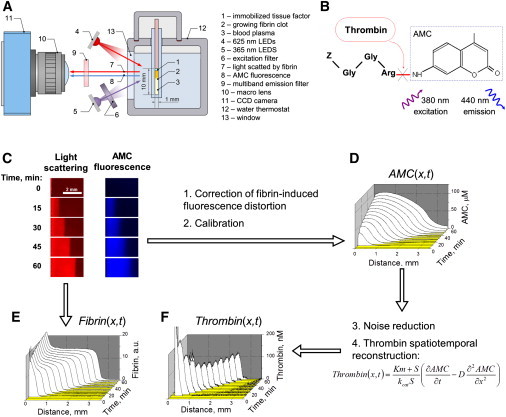
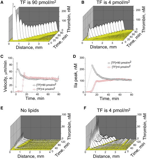
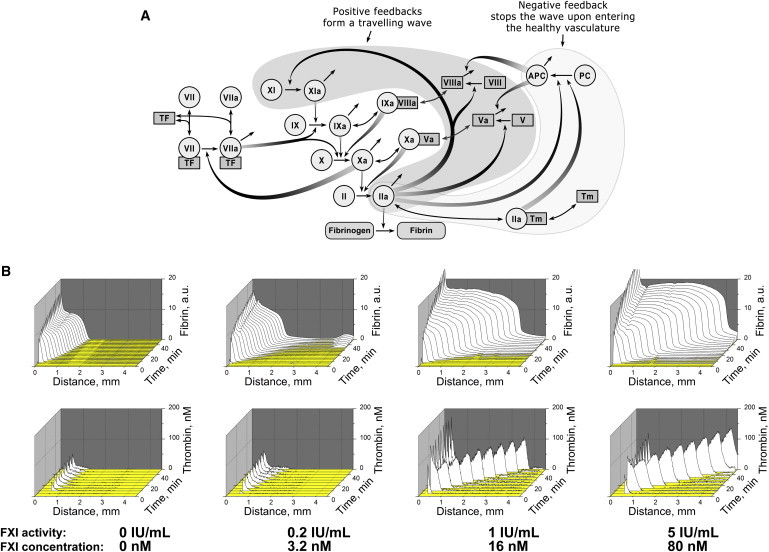
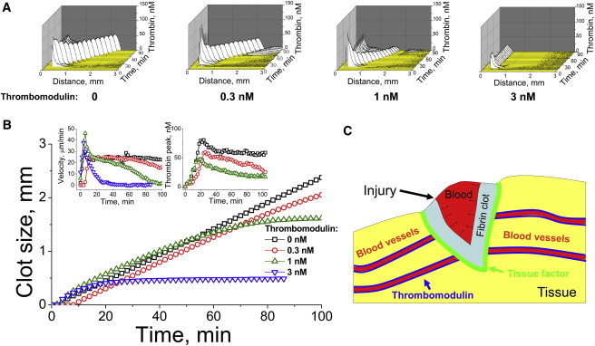
Injury-induced bleeding is stopped by a hemostatic plug formation that is controlled by a complex nonlinear and spatially heterogeneous biochemical network of proteolytic enzymes called blood coagulation. We studied spatial dynamics of thrombin, the central enzyme of this network, by developing a fluorogenic substrate-based method for time- and space-resolved imaging of thrombin enzymatic activity. Clotting stimulation by immobilized tissue factor induced localized thrombin activity impulse that propagated in space and possessed all characteristic traits of a traveling excitation wave: constant spatial velocity, constant amplitude, and insensitivity to the initial stimulation once it exceeded activation threshold. The parameters of this traveling wave were controlled by the availability of phospholipids or platelets, and the wave did not form in plasmas from hemophilia A or C patients who lack factors VIII and XI, which are mediators of the two principal positive feedbacks of coagulation. Stimulation of the negative feedback of the protein C pathway with thrombomodulin produced nonstationary patterns of wave formation followed by deceleration and annihilation. This indicates that blood can function as an excitable medium that conducts traveling waves of coagulation.


Figures

Figure 1
Figure 1
Spatiotemporally resolved imaging of thrombin activity in blood plasma from a healthy individual reveals a propagating wave. (A) Overall experimental design: A layer of immobilized tissue factor (1) induces fibrin clot propagation (2) in nonstirred plasma (3). The sample is illuminated in turn by red (4) or UV LEDs (5) through an excitation filter (6). Light scattered by fibrin (7) and fluorescence of the thrombin-generated AMC (8) pass through a multiband emission filter (9) and macro lens (10) and are recorded by a charge-coupled device (11). (B) Fluorogenic substrate Z-Gly-Gly-Arg-AMC is cleaved by thrombin to yield fluorescent AMC. (C) Time-lapse images: Light scattering from the growing fibrin clot (red) or AMC fluorescence (blue). (D) AMC concentration distribution obtained from fluorescence (C). (E) Fibrin concentration distribution obtained from light scattering (C). (F) Thrombin distribution as a function of space and time obtained from the AMC distribution (E) by solving a reverse reaction-diffusion problem. Plasma is supplemented with 10 μM phospholipids; activation is with 90 pmol TF/m2.
Figure 2
Figure 2
Propagating thrombin wave possesses characteristic properties of a traveling wave in an excitable medium. (A and B) Spatiotemporal thrombin distribution after clotting stimulation in normal plasma supplemented with 10 μM phospholipids with different TF densities. (C and D) Thrombin spatial velocity (A) and peak amplitude (B) for these two experiments. At the p = 0.05 level (n = 8 experiments with plasma from different donors), the thrombin peak height and velocity are not different for these two activation levels. (E and F) Typical thrombin profiles in platelet-free plasma without phospholipid supplementation (E) or in platelet-rich plasma (F). A typical experiment (out of n = 3) is shown. Stimulation is with 4 pmol TF/m2.
Figure 3
Figure 3
Traveling wave of thrombin activity is determined by positive feedbacks in the coagulation network. (A) The coagulation network contains positive feedbacks (filled with gray) that can support self-sustained propagation of the thrombin impulse. Factor XI, located at the top of the cascade, can be activated by thrombin in a feedback that is typical for excitable media. (B) Fibrin clot (upper row) and thrombin formation (lower) in factor XI-deficient plasma supplemented with 5 μM phospholipids and different concentrations of factor XI; a typical experiment is shown (out of n = 2). See also Figs. S5–S8.
Figure 4
Figure 4
Traveling wave propagation is stopped by thrombomodulin: possible implications for confining a hemostatic plug to the wound. (A) Spatiotemporally resolved distribution of thrombin in normal plasma supplemented with different concentrations of thrombomodulin, as indicated in the panels. Plasma is supplemented with 10 μM phospholipids. Activation is with 4 pmol TF/m2. A typical experiment (out of n = 5) is shown. (B) Clot size (main plot), spatial velocity (left inset), and thrombin peak amplitude (right inset) as functions of time for different thrombomodulin concentrations. (C) Hypothetical function of the traveling wave of thrombin in vivo. In a sufficiently large wound, it is necessary to turn all of the blood into a gel, and to spread the clot from the TF-containing injury site into the bulk of the blood. This task is possible, because thrombin propagation can be self-sustained due to factor XI feedback activation, which explains the bleeding upon large injuries in hemophilia C. Abundant thrombomodulin in the healthy blood vessel endothelium prevents the traveling wave from entering the healthy vasculature.

Similar articles

Cited by

References

    1. Ataullakhanov F.I., Guria G.T., Volkova R.I. Spatiotemporal dynamics of clotting and pattern formation in human blood. Biochim. Biophys. Acta. 1998;1425:453–468. - PubMed
    1. Zarnitsina V.I., Pokhilko A.V., Ataullakhanov F.I. A mathematical model for the spatio-temporal dynamics of intrinsic pathway of blood coagulation. II. Results. Thromb. Res. 1996;84:333–344. - PubMed
    1. Rinzel J., Keller J.B. Traveling wave solutions of a nerve conduction equation. Biophys. J. 1973;13:1313–1337. - PMC - PubMed
    1. Loose M., Fischer-Friedrich E., Schwille P. Spatial regulators for bacterial cell division self-organize into surface waves in vitro. Science. 2008;320:789–792. - PubMed
    1. Ovanesov M.V., Ananyeva N.M., Saenko E.L. Initiation and propagation of coagulation from tissue factor-bearing cell monolayers to plasma: initiator cells do not regulate spatial growth rate. J. Thromb. Haemost. 2005;3:321–331. - PubMed

Publication types