Review
. 2012 Nov 30;48(4):491-507.
doi: 10.1016/j.molcel.2012.11.006.

Histone lysine methylation dynamics: establishment, regulation, and biological impact

Affiliations
Review

Histone lysine methylation dynamics: establishment, regulation, and biological impact

Joshua C Black et al. Mol Cell. .

Abstract

Histone lysine methylation has emerged as a critical player in the regulation of gene expression, cell cycle, genome stability, and nuclear architecture. Over the past decade, a tremendous amount of progress has led to the characterization of methyl modifications and the lysine methyltransferases (KMTs) and lysine demethylases (KDMs) that regulate them. Here, we review the discovery and characterization of the KMTs and KDMs and the methyl modifications they regulate. We discuss the localization of the KMTs and KDMs as well as the distribution of lysine methylation throughout the genome. We highlight how these data have shaped our view of lysine methylation as a key determinant of complex chromatin states. Finally, we discuss the regulation of KMTs and KDMs by proteasomal degradation, posttranscriptional mechanisms, and metabolic status. We propose key questions for the field and highlight areas that we predict will yield exciting discoveries in the years to come.


Figures

Figure 1
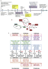
Figure 1. History, Mechanism, and Specificity of KMTs and KDMs
(A) Timeline chronicling important milestones in KMT and KDM research. (B) Schematic depicting generalized reaction mechanisms of KMTs and KDMs. (C) Schematic depicting substrate specificity of KMTs and KDMs.
Figure 2
Figure 2. Distribution of Histone Methylation, KMTs, and KDMs from Genome-wide Profiling Studies
The distribution of methyl modifications is shown relative to chromosomal location, as well as in relationship to active and inactive genes. Green represents euchromatic regions, while red coloring represents heterochromatic regions. Distributions of modifications are represented by bars, and gradients were derived from metagene analysis published as part of the genome-wide data sets from the work of numerous labs. We have included plots from metagene analyses that included both a transcription start site (TSS) and a transcription termination site (TTS). Enzymes marked with an * indicate the distribution depicted from metagene analyses that did not include both a TSS and TTS or from distributions published as heatmaps centered on the TSS. In the active gene model, the green E represents expressed exons, while the red E represents a nonexpressed exon. I denotes introns. Distributions uncovered in specific species are indicated by the following: Y, Saccharomyces cerevisiae; D, Drosophila. The lack of a denotation indicates conservation across multiple species. BV denotes bivalent genes, NBV denotes nonbivalent genes, HOX denotes patterns at Hox genes, and ZNF denotes zinc finger genes. This figure is the compiled work of numerous laboratories. We apologize for being unable to reference everyone’s contributions due to space limitations.
Figure 3
Figure 3. Histone Methylation as Well as KMTs and KDMs Are Dynamically Regulated during the Cell Cycle
Solid shapes denote published information for the levels or functions of the indicated enzymes and modifications during that phase of cell cycle. Histone methylation positions and degree are indicated above the cell-cycle bar, and KMTs and KDMs are indicated below the cell-cycle bar. The white square denotes that the indicated enzyme is important during this time in cell cycle but that expression levels over cell cycle have not been reported. This figure is compiled from work generated by numerous groups. We apologize for being unable to reference everyone’s contributions due to space limitations.
Figure 4
Figure 4. KMTs and KDMs Are Dynamically Regulated through Multiple Mechanisms
KMTs and KDMs are subject to inhibition or activation through the regulatory mechanisms that are indicated.

Similar articles

Cited by

References

    1. Allfrey VG, Mirsky AE. Structural Modifications of Histones and their Possible Role in the Regulation of RNA Synthesis. Science. 1964;144:559. - PubMed
    1. Aoto T, Saitoh N, Sakamoto Y, Watanabe S, Nakao M. Polycomb group protein-associated chromatin is reproduced in post-mitotic G1 phase and is required for S phase progression. J Biol Chem. 2008;283:18905–18915. - PubMed
    1. Baba A, Ohtake F, Okuno Y, Yokota K, Okada M, Imai Y, Ni M, Meyer CA, Igarashi K, Kanno J, et al. PKA-dependent regulation of the histone lysine demethylase complex PHF2-ARID5B. Nat Cell Biol. 2011;13:668–675. - PubMed
    1. Bannister AJ, Kouzarides T. Histone methylation: recognizing the methyl mark. Methods Enzymol. 2004;376:269–288. - PubMed
    1. Barski A, Cuddapah S, Cui K, Roh TY, Schones DE, Wang Z, Wei G, Chepelev I, Zhao K. High-resolution profiling of histone methylations in the human genome. Cell. 2007;129:823–837. - PubMed

Publication types