. 2013 May;34(5):1504-15.
doi: 10.1016/j.neurobiolaging.2012.11.001. Epub 2012 Nov 28.

Adenosine monophosphate-activated protein kinase overactivation leads to accumulation of α-synuclein oligomers and decrease of neurites

Affiliations

Adenosine monophosphate-activated protein kinase overactivation leads to accumulation of α-synuclein oligomers and decrease of neurites

Peizhou Jiang et al. Neurobiol Aging. 2013 May.

Abstract

Neuronal inclusions of α-synuclein (α-syn), termed Lewy bodies, are a hallmark of Parkinson disease (PD). Increased α-syn levels can occur in brains of aging human and neurotoxin-treated mice. Because previous studies have shown increased brain lactate levels in aging brains, in PD affected subjects when compared with age-matched controls, and in mice treated with 1-methyl-4-phenyl-1,2,3,6-tetrahydropyridine(MPTP), we tested the effects of lactate exposure on α-syn in a cell-based study. We demonstrated that (1) lactate treatment led to α-syn accumulation and oligomerization in a time- and concentration-dependent manner; (2) such alterations were mediated via adenosine monophosphate-activated protein kinase (AMPK) and associated with increasing cytoplasmic phosphorylated AMPK levels; (3) AMPK activation facilitated α-syn accumulation and phosphorylation; (4) lactate treatment or overexpression of the active form of AMPK decreased α-syn turnover and neurite outgrowth; and (5) Lewy body-bearing neurons displayed abnormal cytoplasmic distribution of phosphorylated AMPK, which normally is located in nuclei. Together, our results suggest that chronic neuronal accumulation of α-syn induced by lactate-triggered AMPK activation in aging brains might be a novel mechanism underlying α-synucleinopathies in PD and related disorders.


Conflict of interest statement

Disclosure statement

All authors have no actual or potential conflicts of interest. All animal work was approved by the Institutional IACUC.

Figures

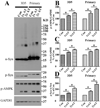
Figure 1
Figure 1. Chronic lactate treatment of 3D5 cells induces AMPK activation and α-syn accumulation in a time- and dose-dependent manner
Cells with retinoic acid elicited neuronal differentiation and 4 days of TetOff induction were treated with (A) 20 mM lactate (LA) for 3 and 6 days or (B) 0 mM, 4 mM, 8 mM, 16 mM and 24 mM lactate for 3 days. Cell lysates were probed with antibodies to α-syn, phosphorylated AMPK (p-AMPK), total-AMPK (T-AMPK) and GAPDH by western blotting. Molecular weight standards and lysates from cells without exposing to lactate were included as references. The results of quantification of α-syn/GAPDH and p-AMPK/T-AMPK immunoreactivities demonstrated in (A), (B) and immunoblots from additional studies were shown in (C) and (D) as percentage of non-treated controls group [i.e. Con0d for (A) and 0 mM for (B)]. In non-treated controls more different sizes α-syn were significantly accumulated in those with longer duration of α-syn overexpression. Lactate treatment caused a significant increase of total α-syn accumulation in a time and concentration manner. Error bars represent standard error of the mean (SEM) (*p<0.05, **P<0.01, n=3). The results are representative of 3 independent experiments.
Figure 2
Figure 2. Chronic lactate treatment leads to AMPK activation and α-syn accumulation in primary neuronal cultures
Lysates from neuronal cultures were probed with antibodies to α-syn, p-AMPK, T-AMPK and GAPDH. The cultures were derived from embryonic (E17) mouse brains, infected with lentivirus carrying Myc-tagged human α-syn for 4 days and followed by 6 and 12 days of exposure to 20 mM lactate, referred to as LA6d and LA12d, respectively. Duplicate cultures with viral infection, but without lactate treatment for 0, 6 and 12 days were included as controls and marked as Con0d, Con6d and Con12d, respectively. Quantitative analyses of immunoreactivities in (A) and immunoblots from additional experiments were performed and the ratio of α-syn/GAPDH and p-AMPK/T-AMPK immunoreactivity were shown in (B) and (C). Bar graphs showed the values of control groups (Con0d) as 100%. Lactate treatment significantly increased the level of total a-syn and that of active AMPK (i.e. p-AMPK). Error bars represent SEM (*p<0.05, **P<0.01, n=3). The results are representative of 3 independent experiments.
Figure 3
Figure 3. Overexpression of AT-AMPK significantly increases AMPK activity (T172 phosphorylated), α-syn accumulation and phosphorylation in 3D5 cells and primary neurons overexpressing human α-syn
3D5 cells and primary neuronal cultures overexpressing α-syn without and with Myc tag, respectively, were infected with 1 arbitrary unit (1x) or 2 units (2x) of lentivirus carrying AT-AMPK and harvested after a 6 days (3D5 cells) and 12 days (primary cultures) of infection. Cells infected with lentivirus carrying empty vector were used as control (Con). Cell lysates were probed with antibodies to total (Synuclein 1) and phosphorylated (p-S129) α-syn, p-AMPK, T-AMPK and GAPDH via western blotting. Results of quantifications of p-AMPK/GAPDH, α-syn/GAPDH and p-syn/α-syn immunoreaectivites in (A) and additional experiments were shown in (B) to (D). Bar graphs showed the values of control groups (Con) as 100%. Cultures overexpressing AT-AMPK contained significantly higher levels of total α-syn and phosphorylated α-syn as well as p-AMPK. These changes are greater in those with its higher doses of viral infection. Error bars represent SEM (*p<0.05, **P<0.01, n=3). The results are representative of 3 independent experiments.
Figure 4
Figure 4. Suppression of AMPK activation inhibits α-syn accumulation and phosphorylation in neuronal cells caused by lactate exposure
(A) Left panel: 3D5 cells differentiated with RA and induced α-syn expression; Right panel: Primary neuronal cultures expressing Myc-tagged human α-syn. The cells were infected with lentivirus carrying empty vector (Con), DN-AMPK (DN) or shRNAs of human or mouse AMPK alpha units (KD) as described in the Method section. Two days post infection 3D5 cells and primary cultures were treated for 3 and 6 days, respectively, with or without 20 mM lactate treatment. Corresponding samples with lactate treatment were referred to as LA, DN/LA and KD/LA, respectively. Cell lysates were probed with antibodies to total and phosphorylated α-syn (p-syn), total (T) and phosphorylated AMPK (p-AMPK) and GAPDH. Results of quantifying the immunoreactivities of T-AMPK/GAPDH, p-AMPK/GAPDH, total α-syn/GAPDH and p-syn/α-syn in (A) and additional experiments were shown in (B) to (E). Bar graphs showed the values of control groups (Con) as 100%. The level of monomeric, total and phosphorylated α-syn as well as that of p-AMPK increased significantly by in LA-treated cultures, but not in those with DN/LA or KD/LA (*p<0.05, **P<0.01, n=3). As expected the level of T-AMPK was significantly less in KD and more in DN cultures than controls. Error bars represent SEM. Asterisk(s) right above the error bar indicated a significant difference between this group and corresponding control (Con). The results are representative of 3 independent experiments.
Figure 5
Figure 5. Interactions between activated AMPK and α-syn
Lysates of cells overexpressing α-syn plus active AMPK (AT-AMPK) were aliquoted. Some were incubated with antibodies to α-syn or p-AMPK to generate immunoprecipitates, regarded as IP/p-syn and IP/p-AMPK, respectively. To verify the specificity of IP some lysates were incubated with mouse or rabbit IgG, and the precipitates obtained were referred to as IP/m IgG and IP/r IgG, respectively. Cell lysates lacking the aforementioned incubation were also included as references. A portion of each aforementioned sample was probed with antibodies to α-syn or p-AMPK to confirm the enrichment of these proteins in IP (A& D), and evaluate their interactions (B & C). Western blotting of IP/α-syn with anti-p-AMPK showed two sizes of p-AMPK immunoreactive proteins of sizes consistent with full-length AMPK (~62 kD) and AT-AMPK (~36 kD) detected in cell lysates. IP/p-AMPK was shown to contain different sizes of α-syn positive proteins corresponding to monomer, dimer and larger oligomer. Arrow heads denote activated endogenous and exogenous AMPK. Arrows denotes both monomeric and dimeric α-syn. Asterisk (*) denotes the heavy chain (55kd) and light chain (25 kd) of IgG in IP. The results are representative of 3 independent experiments.
Figure 6
Figure 6. Activated AMPK phosphorylates α-syn in vitro
Same amount of recombinant α-syn was used for phosphorylation assays of 4 different mixtures, which contain IP/p-AMPK of lysates from BE2-M17D cells expressing (Exp) AT-AMPK (AT) or DN-AMPK (DN), or IP/IgG of lysates from BE2-M17D cells expressing AT-AMPK. ATP was not included in one of the reaction mixtures to serve as a control. Upon the phosphorylation assays were terminated, an equivalent amount of different reaction mixtures was probed for total (top panel in A) and phosphorylated (bottom panel in A) α-syn using antibodies Synuclein 1 and p-S129, respectively. A representative of 3 independent experiments was shown in (A). (B)Quantitative analyses showed the level of α-syn phosphorylation, as evaluated by the ratio of phosphorylated α-syn to total α-syn, differed significantly among different preparations (**P<0.01, n=3 ). Samples containing ATP plus IP/p-AMPK from lysates of AT-AMPK expressing cultures displayed much more phosphorylated α-syn than corresponding IP from lysates of DN-AMPK or IgG precipitates. Phosphorylated α-syn was not readily detected in samples lacking ATP. Error bars represent SEM. Asterisk(s) right above the error bar indicated a significant difference between this group and group 3. The results are representative of 3 independent experiments.
Figure 7
Figure 7. Lactate treatment or overexpressing AT-AMPK causes reduction of neurite outgrowth and suppression of AMPK expression or activation is protective
Representative micrographs showed images of neurons in primary neuronal cultures from PAC wild type α-syn transgenic mice and non-transgenic littermates under different experimental conditions. These include neurons infected with lentivirus carrying (i) empty vector (control), (ii) AT-AMPK (AT), (iii) empty vector plus treatment with 20 mM lactate (LA), (iv) DN-AMPK plus treatment with 20 mM lactate (DN/LA) or (v) shRNA of mouse AMPK alpha units plus exposure to 20 mM lactate (KD/LA). Scale bar: 50 µm. Bar graph shows data summarized from quantitative and statistical analyses of total length of neurites per neuron examined. Error bars represent SEM (*p<0.05). The results are representative of 3 independent experiments.
Figure 8
Figure 8. Lactate treatment or AT-AMPK overexpression decreases α-syn turnover in 3D5 cells without affecting its transcription
(A). Quantitative real-time PCR showed no significant differences (p >0.05, n=3) in the level of α-syn transcripts among 3D5 cells perturbed for 6 days with lactate treatment (LA6d), lentiviral infection of AT-AMPK (AT6d) and lentiviral infection of empty vector (Con6d). Such perturbation was initiated on day 4 of RA-differentiation and TetOff induction, and cells were harvested on day 4 for Con0d and day 10 for the rest. The level of α-syn transcript in non-perturbed cells was significantly (p<0.01, n=3) higher in those harvested with 10 than 4 days of differentiation and induction (Con6d vs Con0d) as expected. LA treatment or expressing AT-AMPK (AT) had no significant effects on the level of α-syn transcript. (B) 3D5 cultures with 4 days of TetOff induction were perturbed with or without lactate treatment (LA), infected with lentiviruses carrying AT-AMPK (also regarded as AT) or empty vector (Con). Three days later, Tet (2µg/ml) was added back to culture media to turn off the expression of exogenous α-syn, and cultures were harvested after 0, 2 and 4 days of the Tet addition. The addition of Tet is known to turn off induced α-syn expression. The lysates of Tet added cultures were probed with antibodies to α-syn and GAPDH as shown in (B). Asterisk (*) indicates a significant difference between marked groups and control. Error bars represent SEM. Analysis of scatter plots between non-degraded α-syn (normalized with GAPDH) levels in different samples and duration of Tet addition was shown in (C). The level of non-degraded α-syn (normalized with GAPDH) in cultures harvested at day 0 was regarded as 100%. The slope (mean±SEM) of extrapolation line obtained from different samples was presented. Statistical analysis showed a significant (*p<0.05, n=3) decrease of α-syn degradation by LA-treatment or AT-AMPK expression when compared to control.
Figure 9
Figure 9. Lactate treatment increases cytoplasmic distribution of activated AMPK in neuronal cells
3D5 cells were induced to express α-syn for 10 days under RA initiated differentiation. On day 4 they were treated with or without 20 mM lactate and referred to as LA or Con, respectively. They were used for dual immunofluorescence labeling with antibodies to p-AMPK and α-syn, followed by staining with DAPI. Panel (A) showed increase of p-AMPK immunolabeling in cytoplasm of the LA-treated. The treatment has very little effects on α-syn distribution. scale bar: 10 µm. Panel (B) showed western blotting of cytoplasmic and nuclear fractions from LA-treated and non-treated controls, using antibodies to p-AMPK, T-AMPK, α-Syn, PARP and HSP90. PARP and HSP90 immunoreactivities were used to check the purity of nuclear and cytoplasm fraction, respectively. Panels (C) to (E) showed the results of quantitative and statistical analyses of data from 3 independent experiments. Cytoplasmic fraction from lactate treated cultures has a significantly higher ratio of p-AMPK to T-AMPK or α-syn to HSP60 (**P<0.01, n=3). In contrast, LA treatment had no significant effects on the ratio of p-AMPK to T-AMPK or α-syn to PARP in nuclear fraction, or the level of T-AMPK to PARP (nuclear) or HSP90 (cytoplasm). Error bars represent SEM. Asterisk (s) right above the error bar indicated a significant difference between this group and its corresponding control (Con). The results are representative of 3 independent experiments.
Figure 10
Figure 10. Subcellular distribution of activated AMPK differs between neurons with and without bearing Lewy bodies
Human specimens containing Basal nucleus of Meynert from different cases with Diffuse Lewy body disease (DLBD) were probed with antibodies to p-AMPK and α-syn by immunocytochemistry and immunofluoresence. In LB-bearing neurons, p-AMPK immunoreactivities were located at the rim of LBs (Left panel in A & upper panel in B) and the rest of neuronal cell bodies. In neurons from basal nucleus of age-matched normal controls, p-AMPK immunoreactivities located mainly in nuclei (Right panel in A & lower panel in B). Arrow head or red label denotes LBs; arrows or green label denotes p-AMPK immunolabeled structures. Scale bar: 20 µm in A and 2 µm in B.

References

    1. Andersen AH, Zhang Z, Zhang M, Gash DM, Avison MJ. Age-associated changes in rhesus CNS composition identified by MRI. Brain Res. 1999;829:90–98. - PubMed
    1. Bowen BC, Block RE, Sanchez-Ramos J, Pattany PM, Lampman DA, Murdoch JB, Quencer RM. Proton MR spectroscopy of the brain in 14 patients with Parkinson disease. AJNR Am J Neuroradiol. 1995;16(1):61–68. - PMC - PubMed
    1. Burré J, Sharma M, Tsetsenis T, Buchman V, Etherton MR, Südhof TC. Alpha-synuclein promotes SNARE-complex assembly in vivo and in vitro. Science. 2010;329(5999):1663–1667. - PMC - PubMed
    1. Calne DB, Langston JW. Aetiology of Parkinson's disease. Lancet. 1983;2(8365-66):1457–1459. - PubMed
    1. Chartier-Harlin MC, Kachergus J, Roumier C, Mouroux V, Douay X, Lincoln S, Levecque C, Larvor L, Andrieux J, Hulihan M, Waucquier N, Defebvre L, Amouyel P, Farrer M, Destée A. Alpha-synuclein locus duplication as a cause of familial Parkinson's disease. Lancet. 2004;364(9440):1167–1169. - PubMed

Publication types