. 2012 Dec 27;2(6):1762-73.
doi: 10.1016/j.celrep.2012.10.026. Epub 2012 Nov 29.

Tracing conidial fate and measuring host cell antifungal activity using a reporter of microbial viability in the lung

Affiliations

Tracing conidial fate and measuring host cell antifungal activity using a reporter of microbial viability in the lung

Anupam Jhingran et al. Cell Rep. .

Abstract

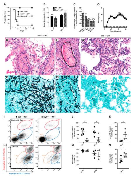
Fluorescence can be harnessed to monitor microbial fate and to investigate functional outcomes of individual microbial cell-host cell encounters at portals of entry in native tissue environments. We illustrate this concept by introducing fluorescent Aspergillus reporter (FLARE) conidia that simultaneously report phagocytic uptake and fungal viability during cellular interactions with the murine respiratory innate immune system. Our studies using FLARE conidia reveal stepwise and cell-type-specific requirements for CARD9 and Syk, transducers of C-type lectin receptor and integrin signals, in neutrophil recruitment, conidial uptake, and conidial killing in the lung. By achieving single-event resolution in defined leukocyte populations, the FLARE method enables host cell profiling on the basis of pathogen uptake and killing and may be extended to other pathogens in diverse model host organisms to query molecular, cellular, and pharmacologic mechanisms that shape host-microbe interactions.


Figures

Figure 1
Figure 1. FLARE Conidia Report Conidial Uptake and Killing In Vitro and In Vivo
(Ai) Fluorescence and (Aii) transmitted light micrographs of (1) Af293, (2) Af293-AF633, (3) Af293-dsRed, and (4) Af293-dsRed-AF633 (FLARE) conidia. (B) The graph shows the average (±SEM) BMM TNF release following coincubation with Af293 (black bar), Af293-dsRed (dark-gray bar), Af293-AF633 (light-gray bar), and FLARE (white bar) conidia at a multiplicity of infection (moi) of 5 (four replicates per strain). ns, not significant. (C) The graph shows dsRed (red) and AF633 (blue) fluorescence and cfu (black circles) from FLARE conidia exposed to the indicated H2O2 concentration. Mean fluorescence intensity is indicated relative to FLARE conidia not treated with H2O2. (D) Scheme of fluorescence changes associated with leukocyte conidial uptake and killing. Bystander leukocytes acquire dsRed and AF633 fluorescence after engulfing FLARE conidia. Loss of dsRed and retention of AF633 fluorescence coincide with conidial killing. (E–G) The plots show lung neutrophils from (E) FLARE-infected, (F) Af293-AF633-infected, or (G) Af293-dsRed-infected mice (inoculum: 3 × 10⁷ conidia) analyzed for dsRed and AF633 fluorescence 36 hr p.i. (H) The graph shows fungal cfu (per sorted event) recovered from sorted dsRed+AF633+ (red bars) or dsRedAF633+ (blue bars) lung neutrophils (Neutr.), macrophages (Macroph.), and CD11b+ DCs 36 hr p.i. (I–M) Fluorescence microscopy (I and J) and imaging cytometry (K–M) of BAL neutrophils from FLARE-infected mice. The micrographs depict sorted dsRed+AF633+ (I and L) and dsRedAF633+ (J and M) and dsRedAF633 neutrophils (K). The columns in (K–M) show bright-field (BF), DAPI, Ly6G, dsRed, and AF633 channels, with composite micrographs (BF, dsRed, and AF633) at the far right. Data are representative from 1 (B), 2 (C), >20 (E–G), or 3 (H–J) experiments.
Figure 2
Figure 2. FLARE Conidia Undergo Leukocyte-Dependent Killing in the Lung
C57BL/6 mice were infected with 3 × 10⁷ FLARE conidia, and single-cell lung suspensions were stained for CD45 and analyzed for dsRed and AF633 fluorescence. (A) Representative plots show live (R1; red gate) and killed conidia (R2; blue gate) in the CD45 or in the CD45+ gate at 36 hr p.i. (B) The bar graphs show the average (±SEM) conidial viability in the CD45 and CD45+ gates (n = 5 per group). *p < 0.05 (Mann-Whitney U test). (C–E) The bar graphs show the average number (±SEM) of (C) leukocytes, (D) fungus-engaged leukocytes, and (E) leukocytes containing killed conidia in FLARE-infected lungs 36 hr p.i. Neutrophils are indicated in black bars, monocytes in dark-gray bars, CD11b+ DCs in gray bars, lung macrophages in light-gray bars, and AMs in white bars. The data in (A–E) are from a representative experiment from more than ten experiments performed with five mice per time point.
Figure 3
Figure 3. NADPH Oxidase Mediates Neutrophil-Dependent Conidial Killing in the Lung
BM chimeric mice (1:1 mix of CD45.1+p47phox(+/+) and CD45.2+p47phox(−/−) BM cells → irradiated CD45.1+CD45.2+ recipients) were infected with 3 × 10⁷ FLARE conidia. (A) Representative plots of p47phox(+/+) and p47phox(−/−) neutrophils analyzed for dsRed and AF633 fluorescence show the frequencies of neutrophils that contain live (red gate) or killed (blue gate) conidia 36 hr p.i. (B and C) Scatterplots from one (36 hr p.i.) or two (12 hr p.i.) experiments show the average frequency (±SEM) of (B) lung neutrophil conidial uptake (R1 + R2) and (C) lung neutrophil conidial viability (R1/(R1 + R2)) by p47phox(+/+) (circles) or p47phox(−/−) (diamonds) neutrophils. *p < 0.05 (paired t test).
Figure 4
Figure 4. CARD9 and Neutrophil Function during Respiratory Fungal Infection
WT (black circles or bars), Dectin-1(−/−) (gray circles or bars), and CARD9(−/−) (white circles or bars) mice were infected via the i.t. route with 8 × 10⁷ Af293 (A and J–M) or 3 × 10⁷ FLARE conidia (B–H and N). (A) Kaplan-Meier survival of WT (n = 8), Dectin-1(−/−) (n = 6), and CARD9(−/−) (n = 6) mice from a representative experiment. (B) Lung cytokines 36 hr p.i. expressed as the fold change (±SEM) in the (−/−) response compared to the (+/+) response pooled from four experiments. (C) Neutrophil recruitment (±SEM) from 8–14 mice, pooled from two to three experiments per time point. (D) Flow cytometry plots of lung neutrophils from FLARE-infected WT, Dectin-1(−/−), and CARD9(−/−) mice. The tan gates indicate bystander neutrophils, and the red (R1) and blue (R2) gates indicate neutrophils that contain live or killed conidia, respectively. (E and G) The plots show neutrophil conidial uptake (R1 + R2) and conidial viability ((R1/(R1 + R2)) (±SEM) in WT, Dectin-1(−/−), and CARD9(−/−) mice at 12, 36, and 72 hr p.i. from a representative experiment. (F and H) The plots show the mean (±SEM) neutrophil conidial uptake and viability using normalized data pooled from four (12 hr), three (36 hr), and two (72 hr) experiments, each with four to five mice per group and time point. (I) ROS generation by BM neutrophils isolated from WT and CARD9(−/−) mice and stimulated with swollen conidia. Data are expressed as RLU/s (×103) (mean ± SEM). (J–L) Representative images of silver-stained lung sections (40×) from WT (J), Dectin-1(−/−) (K), and CARD9(−/−) (L) mice at 3 days p.i. (M) The graph shows the average frequency (±SEM) of germlings in murine lungs 3 days p.i. indicated by arrows in (L). A total of 5,000–7,000 fungal elements were enumerated using sections from two mice per genotype. (N) The graph shows the average (±SEM) lung fungal burden 6 days p.i. The data are pooled from two experiments, each with four to five mice per group. *p < 0.05 (one-way ANOVA). See also Figures S1, S2, S3, and S4.
Figure 5
Figure 5. Syk and Neutrophil Function during Respiratory Fungal Infection
(A–N) Syk(−/−) → WT (hatched circles or bars) and WT → WT (black circles or bars) mice were infected with (A and D–H) Af293 or (B, C, and I–N) FLARE conidia and euthanized at the indicated times p.i. (A) Kaplan-Meier survival plot of Syk(−/−) → WT (n = 10), Dectin-1(−/−)→ WT (n = 5), and WT → WT (n = 10) mice challenged with 2–5 × 10⁷ conidia. The data are pooled from two experiments. (B and C) The bar graphs show the average (±SEM) (B) lung neutrophil recruitment and (C) lung cytokines at 36 hr p.i. Lung cytokine data are expressed as the fold change in the (−/−) response compared to the (+/+) response pooled from two experiments, each with three to four mice per group. (D–H) Representative micrographs of H&E (D, Fi, and G) and silver-stained (E, Fii, and H) lung sections from Syk(−/−) → WT (D, E, Fi, and Fii) and WT → WT (G and H) mice (40×). The dotted lines (black in Fi and white in Fii) show a blood vessel infiltrated by hyphae. (I and L) Representative flow cytometry plots are gated on WT (left column) or Syk(−/−) (right column) (I) lung neutrophils or (L) AMs and analyzed as described in Figure 4. (J, K, M, and N) The scatterplots indicate the mean (±SEM) (J and M) conidial uptake and (K and N) conidial viability in Syk(−/−) and WT, (J and K) neutrophils, and (M and N) AMs. Data from one of two experiments are shown. (O) ROS generation by BM neutrophils isolated from WT → WT and Syk(−/−) → WT mice and stimulated with swollen conidia. Data are expressed as RLU/s (×103) (mean ± SEM). *p < 0.05 (Mann-Whitney U test). See also Figure S5.

Similar articles

Cited by

References

    1. Balajee SA, Marr KA. Conidial viability assay for rapid susceptibility testing of Aspergillus species. J. Clin. Microbiol. 2002;40:2741–2745. - PMC - PubMed
    1. Bonnett CR, Cornish EJ, Harmsen AG, Burritt JB. Early neutrophil recruitment and aggregation in the murine lung inhibit germination of Aspergillus fumigatus Conidia. Infect. Immun. 2006;74:6528–6539. - PMC - PubMed
    1. Boyle KB, Gyori D, Sindrilaru A, Scharffetter-Kochanek K, Taylor PR, Mócsai A, Stephens LR, Hawkins PT. Class IA phosphoinositide 3-kinase β and δ regulate neutrophil oxidase activation in response to Aspergillus fumigatus hyphae. J. Immunol. 2011;186:2978–2989. - PubMed
    1. Brothers KM, Newman ZR, Wheeler RT. Live imaging of disseminated candidiasis in zebrafish reveals role of phagocyte oxidase in limiting filamentous growth. Eukaryot. Cell. 2011;10:932–944. - PMC - PubMed
    1. Brown GD. Innate antifungal immunity: the key role of phagocytes. Annu. Rev. Immunol. 2011;29:1–21. - PMC - PubMed

Publication types

MeSH terms

Substances