

A novel model of spontaneous otitis media with effusion (OME) in the Oxgr1 knock-out mouse
- PMID: 23200873
- PMCID: PMC3535456
- DOI: 10.1016/j.ijporl.2012.09.037
A novel model of spontaneous otitis media with effusion (OME) in the Oxgr1 knock-out mouse
Abstract
Objective: A novel mouse model with a specific genetic mutation in a G protein coupled receptor (GPCR) encoded by the Oxgr1 gene results in a predisposition to spontaneous otitis media with effusion. As a primary component of interest in OME, mucin expression was examined in this model to assess expression as compared to wild type animals and suitability as a murine model of OME.
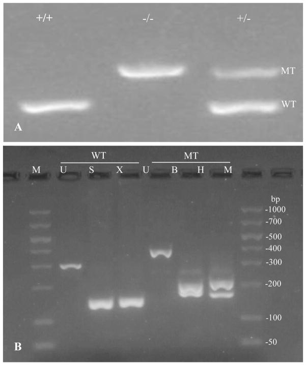
Method: Mutant (Oxgr1(-/-)) and wild-type (Oxgr1(+/+)) mice between ages of 2 and 5 months were examined by otoscopy and auditory brainstem response (ABR). Histology changes in the middle ear were evaluated. Expression of mucin genes in the middle ear epithelium was determined using RT-PCR and quantitative PCR.
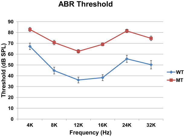
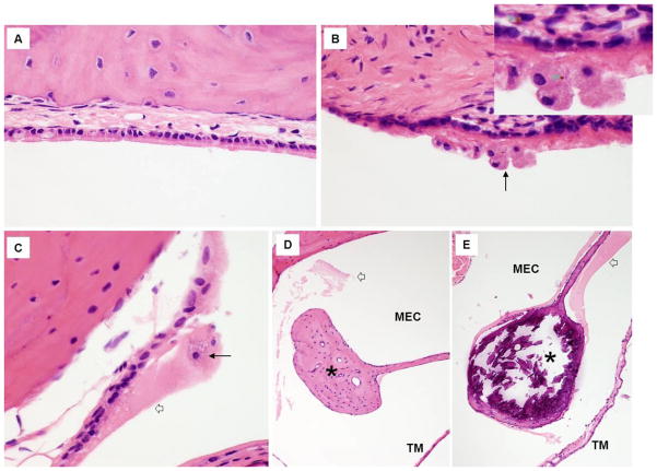
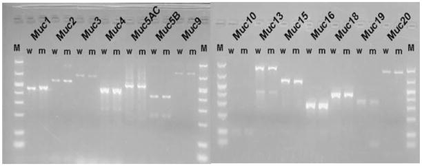
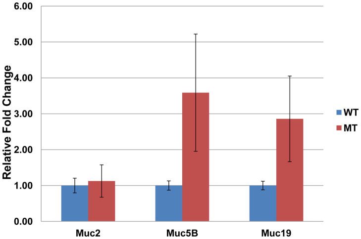
Result: Otoscopic exam showed signs of inflammation in 82% of mutant mice. Significant elevated ABR thresholds were detected in mutant mice indicating hearing loss. Histology analysis of the middle ears demonstrated the presence of inflammatory cells, changes in the mucosal epithelium, and middle ear fluid. RT PCR using universal primers for bacterial 18s rRNA suggested the absence of bacteria in the middle ear. The knockout mice demonstrated expression of Muc1, Muc2, Muc3, Muc4, Muc5AC, Muc5B, Muc9, Muc10, Muc13, Muc15, Muc16, Muc18, Muc19 and Muc20. There was a trend of increase in Muc5B and Muc19 expression in the middle ear of the knockout mice compared to that of wild-type. There was no significant change in the level of Muc2, and Muc5AC was expressed at a level below the detection limit of quantification.
Conclusion: Development of a murine model with genetic defect has several attractive features. The rate of OME in these animals is high at 82%. It is clear that this OME is related to histopathologic changes in the middle ear epithelium of these knock-out mice. Induction of mucus effusion is evident though the viation in dysregulation of GFM does exist in this non-challenge study condition. The underlying cause of these differences between individual animal requires further investigation. Given this, the Oxgr1(-/-) model is likely to be an ideal model to examine mucin regulation in MEE and potentially develop novel GPCR-specific targeted interventions to regulate these processes.
Copyright © 2012 Elsevier Ireland Ltd. All rights reserved.
Conflict of interest statement
None of the authors of this manuscript have any financial or non-financial competing interests to disclose.
Figures
Similar articles
-
Mucin gene expression and mouse middle ear epithelium.Int J Pediatr Otorhinolaryngol. 2010 Aug;74(8):864-8. doi: 10.1016/j.ijporl.2010.04.014. Int J Pediatr Otorhinolaryngol. 2010. PMID: 20846498 Free PMC article.
-
Mucin gene expression in human middle ear epithelium.Laryngoscope. 2007 Sep;117(9):1666-76. doi: 10.1097/MLG.0b013e31806db531. Laryngoscope. 2007. PMID: 17667140
-
Up-regulation of MUC5AC and MUC5B mucin genes in nasopharyngeal respiratory mucosa and selective up-regulation of MUC5B in middle ear in pediatric otitis media with effusion.Laryngoscope. 2006 Mar;116(3):365-9. doi: 10.1097/01.MLG.0000195290.71090.A1. Laryngoscope. 2006. PMID: 16540890
-
Otitis media with effusion.Pediatrics. 2004 May;113(5):1412-29. doi: 10.1542/peds.113.5.1412. Pediatrics. 2004. PMID: 15121966 Review.
-
Mucin gene expression in the rat middle ear: an improved method for RNA harvest.Ann Otol Rhinol Laryngol. 1999 Aug;108(8):762-8. doi: 10.1177/000348949910800809. Ann Otol Rhinol Laryngol. 1999. PMID: 10453784 Review.
Cited by
-
The immunometabolite itaconate stimulates OXGR1 to promote mucociliary clearance during the pulmonary innate immune response.J Clin Invest. 2023 Mar 15;133(6):e160463. doi: 10.1172/JCI160463. J Clin Invest. 2023. PMID: 36919698 Free PMC article.
-
Development of animal models of otitis media.Korean J Audiol. 2013 Apr;17(1):9-12. doi: 10.7874/kja.2013.17.1.9. Epub 2013 Apr 16. Korean J Audiol. 2013. PMID: 24653896 Free PMC article. Review.
-
The transcriptional landscape of the cultured murine middle ear epithelium in vitro.Biol Open. 2021 Apr 15;10(4):bio056564. doi: 10.1242/bio.056564. Epub 2021 Apr 23. Biol Open. 2021. PMID: 33913472 Free PMC article.
-
STAT1 deficiency predisposes to spontaneous otitis media.PLoS One. 2020 Sep 29;15(9):e0239952. doi: 10.1371/journal.pone.0239952. eCollection 2020. PLoS One. 2020. PMID: 32991625 Free PMC article.
-
Development of an In Vivo Model for Eustachian Tube Dysfunction.Bioengineering (Basel). 2022 Jul 15;9(7):317. doi: 10.3390/bioengineering9070317. Bioengineering (Basel). 2022. PMID: 35877368 Free PMC article.
References
-
- Kerschner JE. Mucin gene expression in human middle ear epithelium. Laryngoscope. 2007 Sep;117(9):1666–76. - PubMed
-
- Kerschner JE, Meyer TK, Burrows A. Chinchilla middle ear epithelial mucin gene expression in response to inflammatory cytokines. Arch Otolaryngol Head Neck Surg. 2004 Oct;130(10):1163–7. - PubMed
-
- Kerschner JE, Yang C, Burrows A, Cioffi JA. Signaling pathways in interleukin-1beta-mediated middle ear mucin secretion. Laryngoscope. 2006 Feb;116(2):207–11. - PubMed
-
- Smirnova MG, Kiselev SL, Gnuchev NV, Birchall JP, Pearson JP. Role of the pro-inflammatory cytokines tumor necrosis factor-alpha, interleukin-1 beta, interleukin-6 and interleukin-8 in the pathogenesis of the otitis media with effusion. Eur Cytokine Netw. 2002 Apr-Jun;13(2):161–72. - PubMed
Publication types
MeSH terms
Substances
Grants and funding
LinkOut - more resources
Full Text Sources
Other Literature Sources
Molecular Biology Databases
Research Materials
Miscellaneous
