. 2013 Mar 1;97(3):571-9.
doi: 10.1093/cvr/cvs352. Epub 2012 Dec 1.

SIRT6 protects human endothelial cells from DNA damage, telomere dysfunction, and senescence

Affiliations

SIRT6 protects human endothelial cells from DNA damage, telomere dysfunction, and senescence

Anna Cardus et al. Cardiovasc Res. .

Abstract

Aims: Although endothelial cell senescence is known to play an important role in the development of cardiovascular pathologies, mechanisms that attenuate this process have not been extensively investigated. The aim of this study was to investigate whether SIRT6, a member of the sirtuin family of NAD(+)-dependent protein deacetylases/ADP-ribosyltransferases, protects endothelial cells from premature senescence and dysfunction, and if so which is its mode of action.

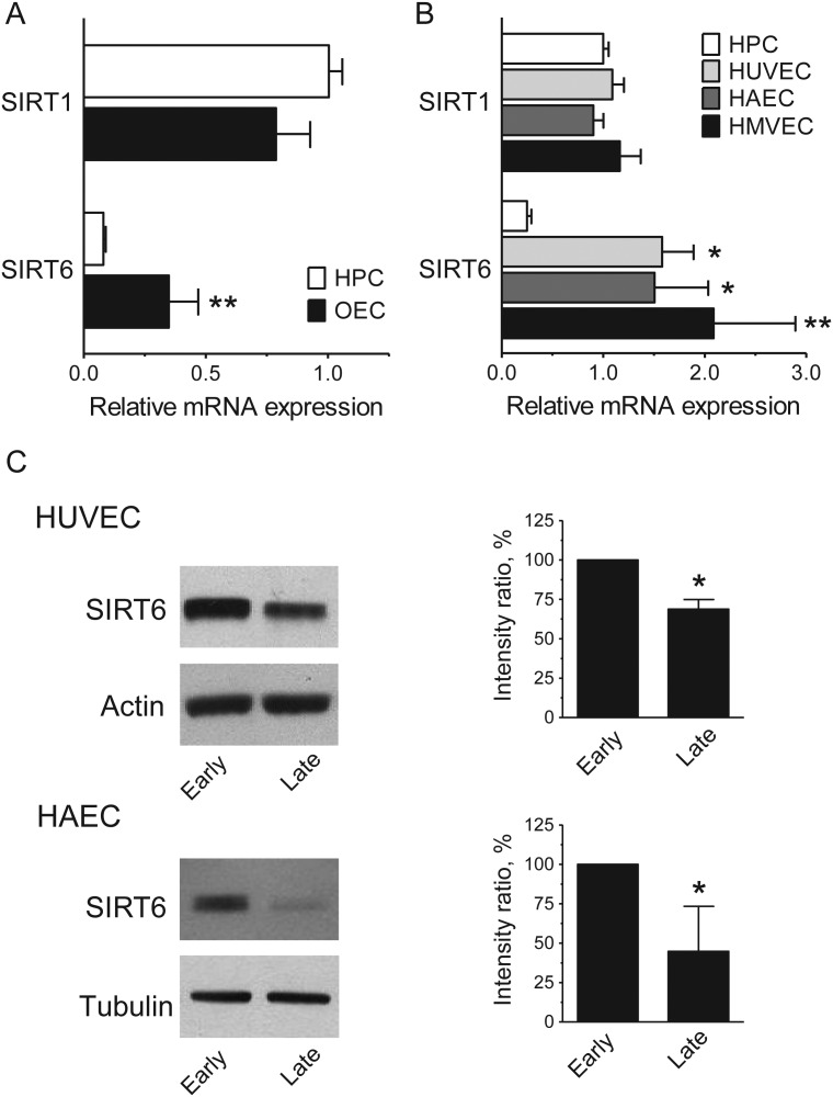
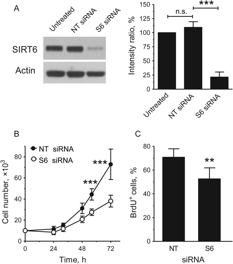
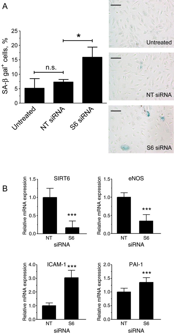
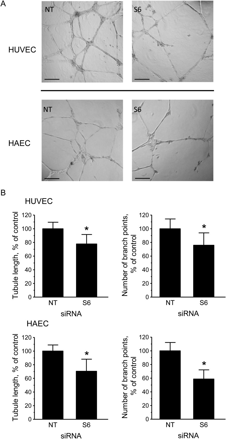
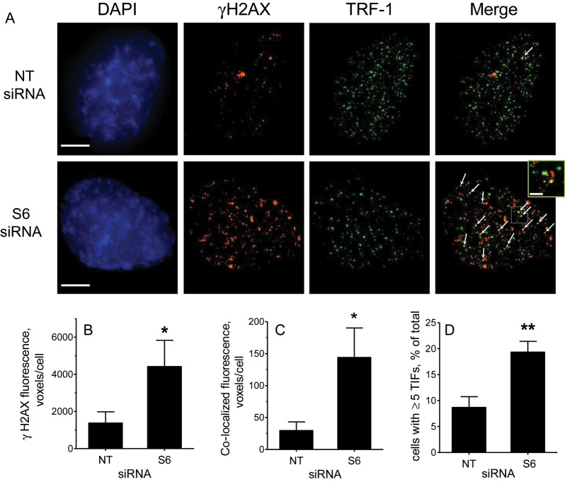
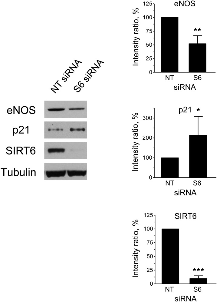
Methods and results: mRNA expression analysis demonstrated comparable levels of SIRT1 and SIRT6 transcripts in endothelial cells derived from different vascular beds and significantly higher levels of SIRT6 in these cells relative to those in haematopoietic progenitor cells. SIRT6 depletion by RNA interference in human umbilical vein endothelial cells (HUVEC) and aortic endothelial cells reduced cell proliferation, increased the fraction of senescence-associated-β-galactosidase-positive cells, and diminished the ability of the cells to form tubule networks on Matrigel. Further examination of SIRT6-depleted HUVEC demonstrated higher intercellular-adhesion molecule-1 (ICAM-1) and plasminogen-activator inhibitor-1 mRNA, lower levels of endothelial nitric oxide synthase mRNA and protein, higher ICAM-1 surface expression, and up-regulation of p21. Fluorescence microscopy of SIRT6-depleted HUVEC stained with anti-phospho-histone H2A.X and anti-telomere-repeat-binding-factor-1 antibodies showed evidence of increased nuclear DNA damage and the formation of telomere dysfunction-induced foci.

Conclusion: This work demonstrates that the presence of SIRT6 in endothelial cells confers protection from telomere and genomic DNA damage, thus preventing a decrease in replicative capacity and the onset of premature senescence. These findings suggest that SIRT6 may be important to maintain endothelial homeostatic functions and delay vascular ageing.


Figures

Figure 1
Figure 1
Expression of SIRT6 in different endothelial cell types. (A) and (B) SIRT1 and SIRT6 mRNA expression profiles. Results are expressed relative to SIRT1 levels in HPC. *P < 0.05, **P < 0.01 vs. SIRT6 in HPC; n = 3–6. (C) SIRT6 protein levels in early- and late-passage cells. Representative immunoblots are shown on the left; quantification is shown on the right. Relative SIRT6 levels were calculated as the ratio of the intensity of the SIRT6 bands to the intensity of the corresponding actin or tubulin bands and results are expressed as a percentage of the level of SIRT6 measured in early-passage cells. *P < 0.05; n = 3.
Figure 2
Figure 2
Depletion of SIRT6 inhibits HUVEC replication. (A) Western blot analysis of SIRT6 protein expression in cells left untreated or harvested 24 h after transfection with either NT or S6 siRNAs. A representative immunoblot is shown on the left; quantification is shown on the right; n = 3. Relative SIRT6 levels were calculated as the ratio of the intensity of the SIRT6 bands to the intensity of the corresponding actin bands and results are expressed as a percentage of the level of SIRT6 measured in the untreated samples. (B) Growth curves and (C) BrdU labelling of transfected cultures; n = 3–4. **P < 0.01 and ***P < 0.001 vs. NT siRNA; n.s., non-significant.
Figure 3
Figure 3
Depletion of SIRT6 in HUVEC induces premature senescence. (A) SA-β-gal staining after three serial transfections; quantification by flow cytometry is shown on the left (n = 4). Representative bright-field images of stained cultures are shown on the right. Bars = 100 μm. (B) mRNA expression analysis (n = 9). *P < 0.05 and ***P < 0.001; n.s., non significant.
Figure 4
Figure 4
Depletion of SIRT6 inhibits in vitro angiogenesis. (A) Representative phase-contrast photomicrographs of tubules formed on Matrigel by NT and S6 siRNA-transfected HUVEC and HAEC. Bars = 200 μm. (B) Comparison of tubule parameters (length and branch points) by computer-assisted image analysis. Results are expressed as a percentage of the values measured in the NT controls. *P < 0.05; n = 5–6.
Figure 5
Figure 5
Depletion of SIRT6 promotes DNA damage and telomere dysfunction. (A) Representative deconvolved images of control (NTsiRNA)- and SIRT6 (S6 siRNA)-depleted HUVEC immunostained with antibodies against γH2AX (red) and TRF-1 (green) and counterstained with DAPI (blue). Arrows in the merged images point to sites of co-localization. The right-most small panel shows an enlarged view of the boxed area in the merged image. Bars = 5 μm. (B and C) Quantification of γH2AX foci and TIFs expressed as mean fluorescence voxels/cell. (D) Quantification of TIFs expressed as the percentage of cells with five or more TIFs/cell. Data represent the average of three experiments with >250 cells scored in each case. *P < 0.05, **P < 0.01.
Figure 6
Figure 6
Depletion of SIRT6 affects eNOS and p21 protein levels. Western blot analysis of SIRT6, eNOS, and p21 protein expression in cells transfected with NT or S6 siRNAs. A representative immunoblot is shown on the left, and quantification is shown on the right. Relative protein levels were calculated as the ratio of the intensity of the indicated bands to the intensity of the corresponding tubulin bands and are expressed as a percentage of the values measured in the NT controls. *P < 0.05, **P < 0.01, and ***P < 0.001; n = 4.

Comment in

References

    1. Campisi J, d'Adda di Fagagna F. Cellular senescence: when bad things happen to good cells. Nat Rev Mol Cell Biol. 2007;8:729–740. - PubMed
    1. Erusalimsky JD, Kurz DJ. Cellular senescence in vivo: its relevance in ageing and cardiovascular disease. Exp Gerontol. 2005;40:634–642. - PubMed
    1. Erusalimsky JD. Vascular endothelial senescence: from mechanisms to pathophysiology. J Appl Physiol. 2009;106:326–332. - PMC - PubMed
    1. Nakagawa T, Guarente L. Sirtuins at a glance. J Cell Sci. 2011;124:833–838. - PMC - PubMed
    1. Ota H, Akishita M, Eto M, Iijima K, Kaneki M, Ouchi Y. Sirt1 modulates premature senescence-like phenotype in human endothelial cells. J Mol Cell Cardiol. 2007;43:571–579. - PubMed

Publication types

MeSH terms

Substances