. 2013 Mar 1:232:182-93.
doi: 10.1016/j.neuroscience.2012.11.033. Epub 2012 Nov 29.

Corneal afferents differentially target thalamic- and parabrachial-projecting neurons in spinal trigeminal nucleus caudalis

Affiliations

Corneal afferents differentially target thalamic- and parabrachial-projecting neurons in spinal trigeminal nucleus caudalis

S A Aicher et al. Neuroscience. .

Abstract

Dorsal horn neurons send ascending projections to both thalamic nuclei and parabrachial nuclei; these pathways are thought to be critical pathways for central processing of nociceptive information. Afferents from the corneal surface of the eye mediate nociception from this tissue which is susceptible to clinically important pain syndromes. This study examined corneal afferents to the trigeminal dorsal horn and compared inputs to thalamic- and parabrachial-projecting neurons. We used anterograde tracing with cholera toxin B subunit to identify corneal afferent projections to trigeminal dorsal horn, and the retrograde tracer FluoroGold to identify projection neurons. Studies were conducted in adult male Sprague-Dawley rats. Our analysis was conducted at two distinct levels of the trigeminal nucleus caudalis (Vc) which receive corneal afferent projections. We found that corneal afferents project more densely to the rostral pole of Vc than the caudal pole. We also quantified the number of thalamic- and parabrachial-projecting neurons in the regions of Vc that receive corneal afferents. Corneal afferent inputs to both groups of projection neurons were also more abundant in the rostral pole of Vc. Finally, by comparing the frequency of corneal afferent appositions to thalamic- versus parabrachial-projecting neurons, we found that corneal afferents preferentially target parabrachial-projecting neurons in trigeminal dorsal horn. These results suggest that nociceptive pain from the cornea may be primarily mediated by a non-thalamic ascending pathway.


Conflict of interest statement

Conflict of Interest Statement

The authors have no known conflicts of interest.

Figures

Figure 1
Figure 1
Schematic representation of the peripheral and central substrates under study. Corneal afferents projecting to the ventrolateral aspect of the caudal (Vc/C1) and rostral (Vi/Vc) trigeminal dorsal horn were labeled with CTb. Concurrent injections of the retrograde tracer FluoroGold into either the thalamic or parabrachial nuclei were utilized to identify populations of projection neurons in the trigeminal dorsal horn. We assessed the connectivity between CTb-ir corneal afferents and FG-ir projection neurons in ventrolateral trigeminal dorsal horn at both the caudal (Vc/C1) and rostral (Vi/Vc) levels of the spinal trigeminal subnucleus caudalis.
Figure 2
Figure 2
FluoroGold (FG) injection sites into the thalamus (A – D) or parabrachial nuclei (E – H). The top panel indicates the rostrocaudal locations of the illustrations in panels A – H. The FG injections that resulted in successful retrograde labeling in the ventrolateral aspect of the trigeminal dorsal horn are shaded with gray and those that did not are represented as open shapes. The injection outlines include both the core of the injection site and the halo often seen with FG injections; thus the injection site depictions are larger than the core area from which neurons were successfully labeled. The two FG injections near thalamus that did not result in labeling in the trigeminal dorsal horn were located too rostrally, illustrated by the open shapes in panel A at −2.1 mm caudal to Bregma. In all successful thalamic cases, the core of the FG injection site that produced retrograde labeling in Vc included Po, VPM, while some also included VPL. The two FG injections near parabrachial nuclei that did not result in labeling were too dorsal (open shapes, E and F). In all successful PB cases, the core of the FG injection site included the lateral PB (LPB). Scale bars = 1 mm. Representative diagrams are modified from the digital atlas of Paxinos and Watson (Paxinos and Watson, 1998) and are reproduced here with permission from the publisher.
Figure 3
Figure 3
Rostrocaudal distribution of retrogradely labeled parabrachial- (white bars) or thalamic- (gray bars) projecting trigeminal neurons and CTb-ir corneal afferents (red circles) in the ventrolateral aspect of the trigeminal dorsal horn. Two relative peaks of CTb-ir corneal afferents were observed; the most caudal peak is at the Vc/C1 transition area (Vc/C1 in green bar), and a more robust peak is seen rostrally at the Vi/Vc transition area (Vi/Vc in green bar). The rostral peak of CTb-ir corneal afferents coincides with the peak of both the parabrachial-projecting and thalamic-projecting neurons. In contrast, a smaller number of parabrachial-projecting neurons were observed and no thalamic-projecting neurons were present in caudal Vc/C1. Regions corresponding to the rostral and caudal peaks of CTb-ir (green bars) were examined using confocal microscopy to look for appositions between CTb-ir corneal afferents and FG-ir projection neurons (green = areas sampled). Scale bar = 1 mm.
Figure 4
Figure 4
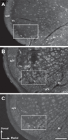
Darkfield micrographs of the ventrolateral trigeminal dorsal horn illustrating retrograde labeling from parabrachial nuclei in caudal (A) and rostral (B) brainstem, and retrograde labeling from the thalamus in rostral brainstem (C). Retrogradely labeled neurons were located primarily in the outer laminae of Vc and at the transition region between Vc and Vi. Retrogradely labeled neurons were also seen in the reticular formation (Rt) but this area was not included in the analysis. The open boxes approximate where cell counts were made as well as where appositions were quantified. The neurons projecting to parabrachial nuclei are more numerous than neurons projecting to thalamic nuclei, both caudally (panel A versus no neurons projecting to thalamus) and rostrally (B versus C). Arrows indicate dorsal and medial directions for orientation on these coronal sections. spV = spinal trigeminal tract. Scale bar = 250 µm.
Figure 5
Figure 5
Corneal afferents form appositions with somata of neurons in the trigeminal dorsal horn. Confocal micrographs of consecutive optical sections in the Z-axis, separated by 0.5 µm, demonstrate a CTb-ir corneal afferent varicosity (white) apposed to a NeuN-ir somata (blue) for two consecutive sections (open arrowheads). Varicose fibers may have multiple contacts with a single neuron, but each varicose fiber was only counted once with respect to a neuronal target for this analysis. Scale bar = 10 µm.
Figure 6
Figure 6
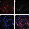
Corneal afferents contact parabrachial-projecting neurons in caudal trigeminal dorsal horn. Confocal micrographs illustrating parabrachial-projecting FG-ir neurons (A, red), CTb-ir corneal afferents (B, white), NeuN-ir somata (C, blue), and the overlay of all three (D) in ventrolateral Vc/C1. Open arrowheads indicate CTb-ir varicosities that are apposed to NeuN-ir only somata. The single closed arrowhead points toward a CTb-ir varicosity that is apposed to a neuron that is immunoreactive for both NeuN as well as FG and are thus parabrachial-projecting. Image is a Z projection of 17 consecutive optical sections for a total thickness of 8.5 µm. Scale bar = 20 µm.
Figure 7
Figure 7
Corneal afferents contact parabrachial-projecting neurons in rostral trigeminal dorsal horn. Confocal micrographs illustrating parabrachial-projecting FG-ir neurons (A, red), CTb-ir corneal afferents (B, white), NeuN-ir somata (C, blue), and the overlay of all three (D) in ventrolateral Vi/Vc. The open arrowhead indicates a CTb-ir varicosity that is apposed to a NeuN-ir only somata. The closed arrowheads indicate CTb-ir varicosities that are apposed to neurons that are immunoreactive for both NeuN as well as FG and are thus parabrachial-projecting. Image is a Z projection of 17 consecutive optical sections for a total thickness of 8.5 µm. Scale bar = 20 µm.
Figure 8
Figure 8
Corneal afferents contact thalamic-projecting neurons in rostral trigeminal dorsal horn. Confocal micrographs illustrating thalamic-projecting FG-ir neurons (A, red), CTb-ir corneal afferents (B, white), NeuN-ir somata (C, blue), and the overlay of all three (D) in the ventrolateral Vi/Vc. Open arrowheads indicate CTb-ir varicosities that are apposed to NeuN-ir only somata. The closed arrowhead indicates a CTb-ir varicosity that is apposed to a neuron that is immunoreactive for both NeuN as well as FG and is thus a thalamic-projecting neuron. Image is a Z projection of 17 consecutive optical sections for a total thickness of 8.5 µm. Scale bar = 20 µm.
Figure 9
Figure 9
The average number of appositions observed between CTb-ir afferent varicosities and somata of neurons at the caudal transition of subnucleus caudalis (Vc/C1; left panel) and the rostral transition of subnucleus caudalis (Vi/Vc; right panel) per animal. Appositions were counted between CTb-ir varicosities and all somata; which included somata displaying immunoreactivity only to the neuronal marker NeuN (black bars), as well as cells that were immunoreactive for both NeuN and FG, the retrograde tracer injected into either thalamic (Thalamic injected, gray bar) or parabrachial nuclei (Parabrachial injected, white bars). Left panel. In Vc/C1, a small number of appositions onto the somata of parabrachial-projecting neurons (Parabrachial Injected; white bar) were observed while no appositions between corneal afferents and thalamic-projecting neurons were observed because no thalamic-projecting neurons were present at this level of the trigeminal subnucleus caudalis (Thalamic Injected). No difference in the average number of CTb-ir afferent appositions onto all neurons (Thalamic Injected vs Parabrachial Injected) was observed (t-test, p=0.43). Right panel. In Vi/Vc, CTb-ir afferents were generally more abundant as compared to Vc/C1, thus there are more CTb-ir afferent appositions onto NeuN-ir only somata (black bars) rostrally (right panel) than caudally (left panel). In Vi/Vc, there were three times more appositions between corneal afferents and parabrachial-projecting neurons (Parabrachial Injected, white bar) than between corneal afferents and somata of thalamic-projecting neurons (Thalamic Injected, gray bar; * Chi-square test, number of CTb-ir varicosities that contact Vi/Vc somata in both sets of FG-injected animals, p < 0.001). No difference in the average number of CTb-ir afferent appositions onto all neurons (Thalamic Injected vs Parabrachial Injected) was observed (t-test, p=0.76).
Figure 10
Figure 10
Schematic representation of the results of the study. More corneal afferents project to the Vi/Vc transition in brainstem (thick arrows), than the Vc/C1 transition area (dashed arrows). A strong direct connection was also observed between CTb-ir corneal afferents in the ventrolateral trigeminal dorsal horn and the somata of parabrachial-projecting neurons, with a more robust connection being seen at the Vi/Vc (thick arrow) transition than the Vc/C1 transition (thin arrow). A small projection was also observed between CTb-ir corneal afferents in the rostral transition zone, Vi/Vc, and the somata of thalamic-projecting nuclei (dashed arrow). There were no thalamic-projecting neurons in ventrolateral Vc/C1, thereby precluding analysis of CTb-ir corneal afferent connectivity to thalamic-projecting somata in this region.

References

    1. Aicher SA, Mitchell JL, Swanson KC, Zadina JE. Endomorphin-2 axon terminals contact mu-opioid receptor-containing dendrites in trigeminal dorsal horn. Brain Res. 2003;977:190–198. - PubMed
    1. Aicher SA, Reis DJ, Nicolae R, Milner TA. Monosynaptic projections from the medullary gigantocellular reticular formation to sympathetic preganglionic neurons in the thoracic spinal cord. J Comp Neurol. 1995;363:563–580. - PubMed
    1. Allen GV, Barbrick B, Esser MJ. Trigeminal-parabrachial connections: Possible pathway for nociception-induced cardiovascular reflex responses. Brain Res. 1996;715:125–135. - PubMed
    1. Bailey TW, Hermes SM, Andresen MC, Aicher SA. Cranial visceral afferent pathways through the nucleus of the solitary tract to caudal ventrolateral medulla or paraventricular hypothalamus: target-specific synaptic reliability and convergence patterns. J Neurosci. 2006;26:11893–11902. - PMC - PubMed
    1. Belmonte C, Acosta MC, Gallar J. Neural basis of sensation in intact and injured corneas. Exp Eye Res. 2004;78:513–525. - PubMed

Publication types

Substances

LinkOut - more resources