

Inter and intra-specific diversity of parasites that cause lymphatic filariasis
- PMID: 23201850
- PMCID: PMC3801268
- DOI: 10.1016/j.meegid.2012.11.002
Inter and intra-specific diversity of parasites that cause lymphatic filariasis
Abstract
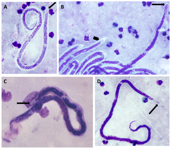
Lymphatic filariasis is caused by three closely related nematode parasites: Wuchereria bancrofti, Brugia malayi and Brugia timori. These species have many ecological variants that differ in several aspects of their biology such as mosquito vector species, host range, periodicity, and morphology. Although the genome of B. malayi (the first genome sequenced from a parasitic nematode) has been available for more than five years, very little is known about genetic variability among the lymphatic dwelling filariae. The genetic diversity among these worms is not only interesting from a biological perspective, but it may have important practical implications for the Global Program to Eliminate Lymphatic Filariasis, as the parasites may respond differently to diagnostic tests and/or medical interventions. Therefore, better information on their genetic variability is urgently needed. With improved methods for nucleic acid extraction and recent advances in sequencing chemistry and instrumentation, this gap can be filled relatively inexpensively. Improved information on filarial genetic diversity may increase the chances of success for lymphatic filariasis elimination programs.
Copyright © 2012 Elsevier B.V. All rights reserved.
Figures
References
-
- Abdullah WO, et al. Detection of circulating antigens and parasite specific antibodies in filariasis. The Southeast Asian journal of tropical medicine and public health. 1993;24(Suppl 2):31–36. - PubMed
-
- Ash LR, Riley JM. Development of subperiodic Brugia malayi in the jird, Meriones unguiculatus, with notes on infections in other rodents. The Journal of parasitology. 1970;56:969–973. - PubMed
-
- Ash LR, Schacher JF. Early life cycle and larval morphogenesis of Wuchereria bancrofti in the jird, Meriones unguiculatus. The Journal of parasitology. 1971;57:1043–1051. - PubMed
-
- Atmosoedjono S, et al. Anopheles balabacensis (Diptera: Culicidae), a vector of Wuchereria kalimantani (Nematoda: Onchocercidae) in east Kalimantan (Borneo), Indonesia. Medical and veterinary entomology. 1993;7:390–392. - PubMed
-
- Bain O, Chabaud AG. Atlas of infective larvae of filariae. Trop Med Parasitol. 1986;37:301–340. - PubMed
Publication types
MeSH terms
Substances
Grants and funding
LinkOut - more resources
Full Text Sources
Other Literature Sources
Miscellaneous
