Sapphire ball lens-based fiber probe for common-path optical coherence tomography and its applications in corneal and retinal imaging
- PMID: 23202062
- PMCID: PMC3534782
- DOI: 10.1364/OL.37.004835
Sapphire ball lens-based fiber probe for common-path optical coherence tomography and its applications in corneal and retinal imaging
Abstract
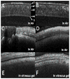
We describe a common-path swept source optical coherence tomography fiber probe design using a sapphire ball lens for cross-sectional imaging and sensing for retina vitrectomy surgery. The high refractive index (n=1.75) of the sapphire ball lens improves the focusing power and enables the probe to operate in the intraocular space. The highly precise spherical shape of the sapphire lens also reduces astigmatism and coma compared to fused nonspherical ball lenses. A theoretical sensitivity model for common-path optical coherence tomography (CP-OCT) was developed to assess its optimal performance based on an unbalanced photodetector configuration. Two probe designs-with working distances 415 and 1221 μm and lateral resolution 11 and 18 μm-were implemented with sensitivity up to 88 dB, which is significantly higher than previously reported CP-OCT probes. We assessed the performances of the fiber probes by cross-sectional imaging a bovine cornea and retina in air and in vitreous gel with a 1310 nm swept source OCT system. To the best of our knowledge, this is the first demonstration of sapphire ball lens-based CP-OCT probes directly inserted into the vitreous gel of a bovine eyeball for ocular imaging with a sensitivity approaching the theoretical limitation of CP-OCT.
Figures


References
-
- Fercher AF, Hitzenberger CK, Kamp G, El-Zaiat SY. Measurement of intraocular distances by backscattering spectral interferometry. Opt Commun. 1995;17:6.
-
- Tearney GJ, Brezinski ME, Bouma BE, Boppart SA, Pitris C, Southern JF, Fujimoto JG. In vivo endoscopic optical biopsy with optical coherence tomography. Science. 1997;276:3. - PubMed
-
- Yamanari M, Makita S, Madjarova VD, Yatagai T, Yasuno Y. Fiber-based polarization-sensitive Fourier domain optical coherence tomography using B-scan-oriented polarization modulation method. Opt Express. 2006;14:6502. - PubMed
Publication types
MeSH terms
Grants and funding
LinkOut - more resources
Full Text Sources
Other Literature Sources
Miscellaneous

