

Modern micro and nanoparticle-based imaging techniques
- PMID: 23202187
- PMCID: PMC3522940
- DOI: 10.3390/s121114792
Modern micro and nanoparticle-based imaging techniques
Abstract
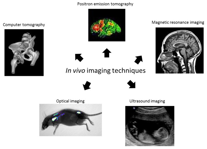
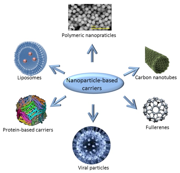
The requirements for early diagnostics as well as effective treatment of insidious diseases such as cancer constantly increase the pressure on development of efficient and reliable methods for targeted drug/gene delivery as well as imaging of the treatment success/failure. One of the most recent approaches covering both the drug delivery as well as the imaging aspects is benefitting from the unique properties of nanomaterials. Therefore a new field called nanomedicine is attracting continuously growing attention. Nanoparticles, including fluorescent semiconductor nanocrystals (quantum dots) and magnetic nanoparticles, have proven their excellent properties for in vivo imaging techniques in a number of modalities such as magnetic resonance and fluorescence imaging, respectively. In this article, we review the main properties and applications of nanoparticles in various in vitro imaging techniques, including microscopy and/or laser breakdown spectroscopy and in vivo methods such as magnetic resonance imaging and/or fluorescence-based imaging. Moreover the advantages of the drug delivery performed by nanocarriers such as iron oxides, gold, biodegradable polymers, dendrimers, lipid based carriers such as liposomes or micelles are also highlighted.
Figures
References
-
- Llinas R.R., Walton K.D., Nakao M., Hunter I., Anquetil P.A. Neuro-vascular central nervous recording/stimulating system: Using nanotechnology probes. J. Nanopart. Res. 2005;7:111–127.
-
- Santra S., Yang H., Stanley J.T., Holloway P.H., Moudgil B.M., Walter G., Mericle R.A. Rapid and effective labeling of brain tissue using TAT-conjugated CdS: Mn/ZnS quantum dots. Chem. Commun. 2005;2005:3144–3146. - PubMed
-
- Keefer E.W., Botterman B.R., Romero M.I., Rossi A.F., Gross G.W. Carbon nanotube coating improves neuronal recordings. Nat. Nanotechnol. 2008;3:434–439. - PubMed
-
- Neuwelt E.A., Varallyay C.G., Manninger S., Solymosi D., Haluska M., Hunt M.A., Nesbit G., Stevens A., Jerosch-Herold M., Jacobs P.M., et al. The potential of ferumoxytol nanoparticle magnetic resonance imaging, perfusion, and angiograpgy in central nervous system malignancy: A pilot study. Neurosurgery. 2007;60:601–611. - PubMed
-
- Neuwelt E.A., Varallyay P., Bago A.G., Muldoon L.L., Nesbit G., Nixon R. Imaging of iron oxide nanoparticles by MR and light microscopy in patients with malignant brain tumours. Neuropathol. Appl. Neurobiol. 2004;30:456–471. - PubMed
Publication types
MeSH terms
LinkOut - more resources
Full Text Sources
Medical
