

Interleukin-17 (IL-17) expression is reduced during acute myocardial infarction: role on chemokine receptor expression in monocytes and their in vitro chemotaxis towards chemokines
- PMID: 23202375
- PMCID: PMC3528254
- DOI: 10.3390/toxins4121427
Interleukin-17 (IL-17) expression is reduced during acute myocardial infarction: role on chemokine receptor expression in monocytes and their in vitro chemotaxis towards chemokines
Abstract
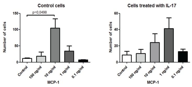
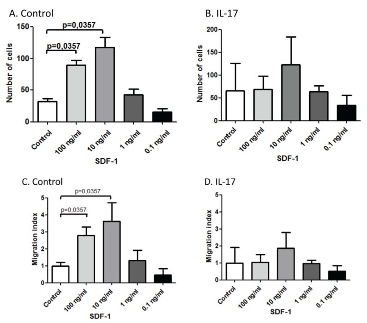
The roles of immune cells and their soluble products during myocardial infarction (MI) are not completely understood. Here, we observed that the percentages of IL-17, but not IL-22, producing cells are reduced in mice splenocytes after developing MI. To correlate this finding with the functional activity of IL-17, we sought to determine its effect on monocytes. In particular, we presumed that this cytokine might affect the chemotaxis of monocytes important for cardiac inflammation and remodeling. We observed that IL-17 tends to reduce the expression of two major chemokine receptors involved in monocyte chemotaxis, namely CCR2 and CXCR4. Further analysis showed that monocytes pretreated with IL-17 have reduced in vitro chemotaxis towards the ligand for CCR2, i.e., MCP-1/CCL2, and the ligand for CXCR4, i.e., SDF-1α/CXCL12. Our results support the possibility that IL-17 may be beneficial in MI, and this could be due to its ability to inhibit the migration of monocytes.
Figures


Similar articles
-
Heterologous desensitization of opioid receptors by chemokines inhibits chemotaxis and enhances the perception of pain.Proc Natl Acad Sci U S A. 2002 Aug 6;99(16):10276-81. doi: 10.1073/pnas.102327699. Epub 2002 Jul 18. Proc Natl Acad Sci U S A. 2002. PMID: 12130663 Free PMC article.
-
Ly6Chigh Monocytes Oscillate in the Heart During Homeostasis and After Myocardial Infarction-Brief Report.Arterioscler Thromb Vasc Biol. 2017 Sep;37(9):1640-1645. doi: 10.1161/ATVBAHA.117.309259. Epub 2017 Jun 29. Arterioscler Thromb Vasc Biol. 2017. PMID: 28663258
-
High-density lipoproteins suppress chemokines and chemokine receptors in vitro and in vivo.Arterioscler Thromb Vasc Biol. 2010 Sep;30(9):1773-8. doi: 10.1161/ATVBAHA.110.211342. Epub 2010 Aug 11. Arterioscler Thromb Vasc Biol. 2010. PMID: 20702809
-
Monocyte subtypes and the CCR2 chemokine receptor in cardiovascular disease.Clin Sci (Lond). 2017 Jun 1;131(12):1215-1224. doi: 10.1042/CS20170009. Clin Sci (Lond). 2017. PMID: 28566450 Review.
-
Role of the SDF-1/CXCR4 system in myocardial infarction.Circ J. 2010 Mar;74(3):418-23. doi: 10.1253/circj.cj-09-1021. Epub 2010 Jan 30. Circ J. 2010. PMID: 20118565 Review.
Cited by
-
Inflammation Markers and Major Depressive Disorder in Patients With Chronic Heart Failure: Results From the Sertraline Against Depression and Heart Disease in Chronic Heart Failure Study.Psychosom Med. 2015 Sep;77(7):808-15. doi: 10.1097/PSY.0000000000000216. Psychosom Med. 2015. PMID: 26186432 Free PMC article.
-
Bioinformatics and Experimental Validation of Diagnostic Marker Genes for Myocardial Infarction and Analysis of Their Immune Cell Infiltration.Biochem Genet. 2025 Aug 19. doi: 10.1007/s10528-025-11211-2. Online ahead of print. Biochem Genet. 2025. PMID: 40828202
-
On the role of natural killer cells in neurodegenerative diseases.Toxins (Basel). 2013 Feb 19;5(2):363-75. doi: 10.3390/toxins5020363. Toxins (Basel). 2013. PMID: 23430541 Free PMC article. Review.
-
Gene expression pattern of TCR repertoire and alteration expression of IL-17A gene of γδ T cells in patients with acute myocardial infarction.J Transl Med. 2018 Jul 5;16(1):189. doi: 10.1186/s12967-018-1567-7. J Transl Med. 2018. PMID: 29976209 Free PMC article.
-
Interleukin-17 and acute coronary syndrome.J Zhejiang Univ Sci B. 2013 Aug;14(8):664-9. doi: 10.1631/jzus.BQICC701. J Zhejiang Univ Sci B. 2013. PMID: 23897784 Free PMC article. Review.
References
-
- Maugeri N., Rovere-Querini P., Evangelista V., Godino C., Demetrio M., Baldini M., Figini F., Coppi G., Slavich M., Camera M., et al. An intense and short-lasting burst of neutrophil activation differentiates early acute myocardial infarction from systemic inflammatory syndromes. PLoS One. 2012;7:e39484. - PMC - PubMed
-
- Imanishi T., kasaka T. Biomarkers associated with vulnerable atheromatous plaque. Curr. Med. Chem. 2012;19:2588–2596. - PubMed
Publication types
MeSH terms
Substances
LinkOut - more resources
Full Text Sources
Medical
Miscellaneous
