A characterization of aerosolized Sudan virus infection in African green monkeys, cynomolgus macaques, and rhesus macaques
- PMID: 23202456
- PMCID: PMC3497044
- DOI: 10.3390/v4102115
A characterization of aerosolized Sudan virus infection in African green monkeys, cynomolgus macaques, and rhesus macaques
Abstract
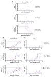
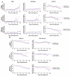
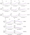
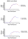
Filoviruses are members of the genera Ebolavirus, Marburgvirus, and "Cuevavirus". Because they cause human disease with high lethality and could potentially be used as a bioweapon, these viruses are classified as CDC Category A Bioterrorism Agents. Filoviruses are relatively stable in aerosols, retain virulence after lyophilization, and can be present on contaminated surfaces for extended periods of time. This study explores the characteristics of aerosolized Sudan virus (SUDV) Boniface in non-human primates (NHP) belonging to three different species. Groups of cynomolgus macaques (cyno), rhesus macaques (rhesus), and African green monkeys (AGM) were challenged with target doses of 50 or 500 plaque-forming units (pfu) of aerosolized SUDV. Exposure to either viral dose resulted in increased body temperatures in all three NHP species beginning on days 4-5 post-exposure. Other clinical findings for all three NHP species included leukocytosis, thrombocytopenia, anorexia, dehydration, and lymphadenopathy. Disease in all of the NHPs was severe beginning on day 6 post-exposure, and all animals except one surviving rhesus macaque were euthanized by day 14. Serum alanine transaminase (ALT) and aspartate transaminase (AST) concentrations were elevated during the course of disease in all three species; however, AGMs had significantly higher ALT and AST concentrations than cynos and rhesus. While all three species had detectable viral load by days 3-4 post exposure, Rhesus had lower average peak viral load than cynos or AGMs. Overall, the results indicate that the disease course after exposure to aerosolized SUDV is similar for all three species of NHP.
Figures


Similar articles
-
Natural history of Sudan ebolavirus infection in rhesus and cynomolgus macaques.Emerg Microbes Infect. 2022 Dec;11(1):1635-1646. doi: 10.1080/22221751.2022.2086072. Emerg Microbes Infect. 2022. PMID: 35657325 Free PMC article.
-
Aerosolized rift valley fever virus causes fatal encephalitis in african green monkeys and common marmosets.J Virol. 2014 Feb;88(4):2235-45. doi: 10.1128/JVI.02341-13. Epub 2013 Dec 11. J Virol. 2014. PMID: 24335307 Free PMC article.
-
Aerosol exposure to Zaire ebolavirus in three nonhuman primate species: differences in disease course and clinical pathology.Microbes Infect. 2011 Oct;13(11):930-6. doi: 10.1016/j.micinf.2011.05.002. Epub 2011 May 25. Microbes Infect. 2011. PMID: 21651988
-
Current perspectives on the phylogeny of Filoviridae.Infect Genet Evol. 2011 Oct;11(7):1514-9. doi: 10.1016/j.meegid.2011.06.017. Epub 2011 Jun 30. Infect Genet Evol. 2011. PMID: 21742058 Free PMC article. Review.
-
Animal models for filovirus infections.Zool Res. 2018 Jan 18;39(1):15-24. doi: 10.24272/j.issn.2095-8137.2017.053. Zool Res. 2018. PMID: 29511141 Free PMC article. Review.
Cited by
-
Natural History of Aerosol-Induced Ebola Virus Disease in Rhesus Macaques.Viruses. 2021 Nov 17;13(11):2297. doi: 10.3390/v13112297. Viruses. 2021. PMID: 34835103 Free PMC article.
-
STAT-1 Knockout Mice as a Model for Wild-Type Sudan Virus (SUDV).Viruses. 2021 Jul 17;13(7):1388. doi: 10.3390/v13071388. Viruses. 2021. PMID: 34372594 Free PMC article.
-
Development of a Well-Characterized Cynomolgus Macaque Model of Sudan Virus Disease for Support of Product Development.Vaccines (Basel). 2022 Oct 15;10(10):1723. doi: 10.3390/vaccines10101723. Vaccines (Basel). 2022. PMID: 36298588 Free PMC article.
-
Oral and Conjunctival Exposure of Nonhuman Primates to Low Doses of Ebola Makona Virus.J Infect Dis. 2016 Oct 15;214(suppl 3):S263-S267. doi: 10.1093/infdis/jiw149. Epub 2016 Jun 9. J Infect Dis. 2016. PMID: 27284090 Free PMC article.
-
Comparison of the Aerosol Stability of 2 Strains of Zaire ebolavirus From the 1976 and 2013 Outbreaks.J Infect Dis. 2016 Oct 15;214(suppl 3):S290-S293. doi: 10.1093/infdis/jiw193. Epub 2016 Aug 8. J Infect Dis. 2016. PMID: 27503365 Free PMC article.
References
-
- Sanchez A., Geisbert T.W., Feldman H. Filoviridae: Marburg and Ebola Viruses. In: Knipe D.M., Howley P.M., editors. Fields Virology. 5th. Lippincott Williams & Wilkins; Philadelphia, USA: 2006. pp. 1409–1448.
-
- Leroy E.M., Kumulungui B., Pourrut X., Rouquet P., Hassanin A., Yaba P., Delicat A., Paweska J.T., Gonzalez J.P., Swanepoel R. Fruit bats as reservoirs of Ebola virus. Nature. 2005;438:575–576. - PubMed
-
- Kuhn J.H., Becker S., Ebihara H., Geisbert T.W., Johnson K.M., Kawaoka Y., Lipkin W.I., Negredo A.I., Netesov S.V., Nichol S.T., et al. Proposal for a revised taxonomy of the family Filoviridae: Classification, names of taxa and viruses, and virus abbreviations. Arch. Virol. 2010;155:2083–2103. doi: 10.1007/s00705-010-0814-x. - DOI - PMC - PubMed
-
- King A.M.Q., Adams M.J., Carstens E.B., Lefkowitz E.J. Virus Taxonomy-Ninth Report of the International Committee on Taxonomy of Viruses. E.A. Press; London, UK: 2011. pp. 665–671.
Publication types
MeSH terms
Substances
LinkOut - more resources
Full Text Sources

