. 2012 Oct 16;4(10):2137-61.
doi: 10.3390/v4102137.

D471G mutation in LCMV-NP affects its ability to self-associate and results in a dominant negative effect in viral RNA synthesis

Affiliations

D471G mutation in LCMV-NP affects its ability to self-associate and results in a dominant negative effect in viral RNA synthesis

Emilio Ortiz-Riaño et al. Viruses. .

Abstract

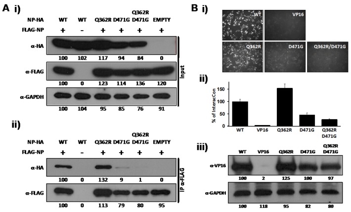
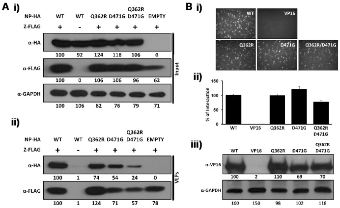
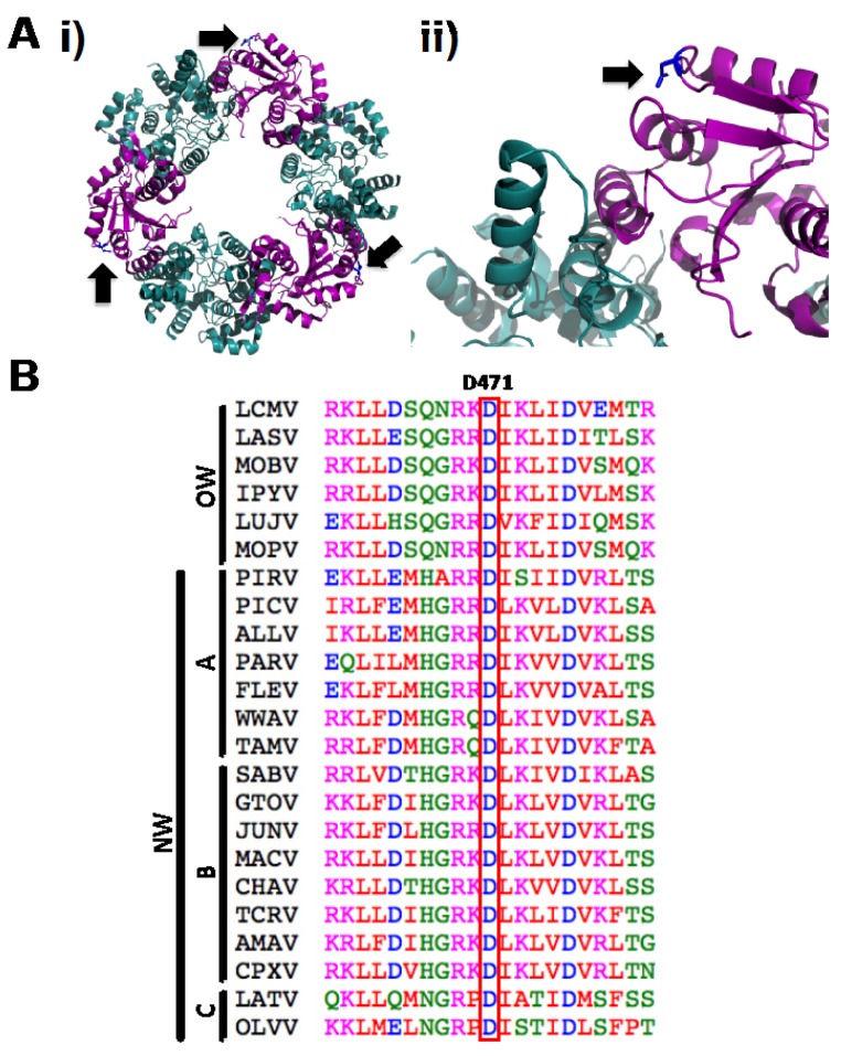
Arenaviruses merit significant interest because several family members are etiological agents of severe hemorrhagic fevers, representing a major burden to public health. Currently, there are no FDA-licensed vaccines against arenaviruses and the only available antiviral therapy is limited to the use of ribavirin that is partially effective. Arenavirus nucleoprotein (NP) is found associated with the genomic RNA forming the viral ribonucleoproteins (vRNPs) that together with the polymerase (L) direct viral replication and transcription. Virion formation requires the recruitment of vRNPs into budding sites, a process in which the arenavirus matrix-like protein (Z) plays a major role. Therefore, proper NP-NP and NP-Z interactions are required for the generation of infectious progeny. In this work we demonstrate the role of the amino acid residue D471 in the self-association of lymphocytic choriomeningitis virus nucleoprotein (LCMV-NP). Amino acid substitutions at this position abrogate NP oligomerization, affecting its ability to mediate replication and transcription of a minigenome reporter plasmid. However, its ability to interact with the Z protein, counteract the cellular interferon response and bind to dsRNA analogs was retained. Additionally, we also document the dominant negative effect of D471G mutation on viral infection, suggesting that NP self-association is an excellent target for the development of new antivirals against arenaviruses.


Figures

Figure 1
Figure 1
LCMV-NP D471G single amino acid mutant is affected in self-association. (A) Co-IP: Human 293T cells were co-transfected with 2 μg of the pC- LCMV-NP wt, single (Q362R and D471G), or double (Q362R/D471G) amino acid mutants HA-tagged, together with 2 μg of pC-LCMV-NP wt FLAG-tagged expression plasmid. As controls, LCMV-NP tagged versions were expressed individually together with 2 μg of empty pC to keep constant the total amount of transfected DNA. At 48 hpt, cell lysates were prepared and analyzed for protein expression levels by WB using anti-HA or anti-FLAG polyclonal antibodies (i). GAPDH was used as a loading control. Cell lysates were immunoprecipitated with anti-FLAG affinity agarose beads (ii) and analyzed by WB with the indicated antibodies (left). Numbers at the bottom of each WB lane represent the quantification of band intensities normalized to the signal of cells co-transfected with HA- and FLAG-tagged wt NPs, as described in Material and Methods. (B) M2H: Human 293T cells were co-transfected in triplicate with 2 μg of the indicated wt, single, or double amino acid mutant pC-VP16-tagged expression plasmids, together with 2 μg of LCMV-NP-GAL4 expression plasmid, along with 1 μg of the pG5 GFP/FFL dual reporter plasmid, and 0.1 μg of the pRL SV40 expression plasmid to normalize transfection efficiencies. At 72 hpt, GFP expression was assessed using fluorescence microscopy and cell extracts were prepared to determine the strength of NP-NP interaction using the Promega dual-luciferase reporter assay and a Lumicount luminometer. As negative control, we transfected cells with pC-NP-GAL4 together with pC-VP16. Representative fields of transfected cells are illustrated (i). Reporter gene activation (FFL) is shown as percentage (%) of LCMV NP-NP wt interaction (pC-NP-VP16 and pC-NP-GAL4) after normalization of transfection efficiencies with the Renilla luciferase expression plasmid pRL SV40 (ii). Cell lysates were used to detect expression of LCMV-NP wt and mutants by WB using an anti-VP16 polyclonal antibody (iii). GAPDH was used as a loading control. Numbers at the bottom of each WB lane represent the quantification of band intensities normalized to wt NP, as described in material and methods.
Figure 2
Figure 2
D471G amino acid substitution in LCMV-NP does not affect its interaction with LCMV-Z. (A) VLP assay: Human 293T cells were co-transfected with 2 μg of the indicated pC wt or mutant NPs HA-tagged, together with 2 μg of pC-LCMV-Z FLAG-tagged expression plasmid. Empty pC plasmid was included to normalize the total amount of transfected DNA. At 72 hpt, cell lysates were prepared and analyzed for protein expression levels for NP (α-HA) and Z (α-FLAG) (i). GAPDH was used as a loading control. TCS from same transfected cells were used for isolation of VLP to evaluate NP incorporation (α-HA) into Z-mediated VLP (α-FLAG) (ii). Numbers at the bottom of each WB lane represent the quantification of band intensities normalized to NP and Z wt expression levels. (B) M2H assay: Human 293T cells (6.5x10⁵) were co-transfected in triplicate (12-well plate format) with 2 μg of the indicated pC-VP16-tagged wt or mutant NPs, together with 2 μg of pC-GAL4-tagged Z expression plasmid as described in Figure 1B. At 72 hpt, LCMV NP-Z interaction was determined by GFP expression (i) and by luciferase activity (ii). pC-VP16 expression plasmid was used as negative control. Reporter gene activation (FFL) is shown as percentage of wt interaction (pC-NP-VP16 and pC-GAL4-Z) after normalization of transfection efficiencies with the Renilla luciferase expression plasmid pRL SV40. Expression levels of wt and mutant NP were determined by WB using an anti-VP16 polyclonal antibody (iii). GAPDH was used as a loading control. Numbers at the bottom of each WB lane represent the quantification of band intensities normalized to wt NP lane as described in material and methods.
Figure 3
Figure 3
D471G substitution affects the ability of LCMV-NP to promote replication and gene expression of an LCMV MG. BHK-21 cells were co-transfected in triplicate with 0.5 μg of pPolI GFP-Pur/Gluc, together with 0.6 μg of pC-L, 0.3 μg of pC wt or mutant NPs HA-tagged, and 0.1μg of the pSV40-Cluc expression vector to normalize transfection efficiencies At 48 hpt, MG driven GFP expression (A) and luciferase activity in TCS (B) were determined. Cell lysates were used to determine expression levels of wt and mutant NPs by WB using an anti-HA antibody (C). GAPDH was used as a loading control. Empty pC was used as a negative control. Percentages of relative luciferase units (% RLUs) were normalized with respect to the activity of the wt NP, after normalization of transfection efficiencies based on Cluc luminescence values. Numbers at the bottom of each WB lane represent the quantification of band intensities normalized to wt NP lane as described in material and methods.
Figure 4
Figure 4
D471G amino acid substitution does not affect the anti-IFN-I function of LCMV-NP. Human 293T cells were co-transfected in triplicate with 0.5 μg of each of the IFNβ reporter plasmids (pIFNβ-GFP and pIFNβ-FFL), 0.1 μg of the indicated pC-NP HA-tagged expression vector, and 0.1 μg of pRL SV40 expression plasmid to normalize transfection efficiencies. At 16 hpt, cells were mock infected or infected with SeV (moi=3) to induce activation of the IFNβ promoter, and 24 hours later, GFP expression was assessed by fluorescence microscopy (A). Cell lysates were prepared for luciferase activities (B), and to detect expression of wt and mutant NPs by WB using an anti-HA antibody (C). GAPDH was used as a loading control. Reporter gene activation is shown as % of RLUs of a SeV-infected, empty-vector transfected control, after normalization by RL luminescence values. Numbers at the bottom of each WB lane represent the quantification of band intensities normalized to wt NP lane, as described in material and methods.
Figure 5
Figure 5
D471G amino acid substitution does not affect dsRNA binding of LCMV-NP. Human 293T cells were co-transfected with 2.5 μg of the indicated pC-LCMV-NP wt or amino acid mutants HA-tagged. pC-LCMV-Z HA-tagged was used as negative control for binding. At 48 hpt, cell lysates were prepared and analyzed for NP expression levels by WB using an anti-HA antibody (A). GAPDH was used as a loading control. Cell lysates were used in pull-down assays with poly I:C bound to sepharose beads and immunoprecipitates analyzed by WB (B). Numbers at the bottom of each WB lane represent the quantification of band intensities normalized to wt NP lane, as described in material and methods.
Figure 6
Figure 6
Effects of D471 substitution on LCMV-NP functions. (A) NP-NP interaction: Human 293T cells were co-transfected as described in Figure 1B with 2 μg of the indicated pC wt or mutant NP-VP16 fusion proteins, together with 2 μg of NP-GAL4 expression plasmids. At 72 hpt, cell extracts were prepared to determine the strength of the interaction. VP16 expression plasmid was used as negative control. Reporter gene activation (FFL) is shown as percentage of wt interaction (pC-NP-VP16 and pC-NP-GAL4) after normalization of transfection efficiencies based on levels of Renilla luciferase activity driven by plasmid pRL SV40. Cell lysates were used to detect expression of wt and mutant NPs by WB using an anti-VP16 polyclonal antibody. GAPDH was used as a loading control. (B) NP-Z interaction: Human 293T cells were co-transfected as described in Figure 2B with 2 μg of the indicated pC wt or mutant NP-VP16, together with 2 μg of GAL4-Z expression plasmids. At 48 hpt, cell extracts were prepared to determine the strength of the interaction and protein expression. Reporter gene activation (FFL) is shown as percentage of wt interaction (pC-NP-VP16 and pC-GAL4-Z) after normalization of transfection efficiencies based on Renilla luciferase values. Cell lysates were used to detect expression of wt and mutant NPs by WB using an anti-VP16 polyclonal antibody. GAPDH was used as a loading control. (C) Replication and transcription activity: BHK-21 cells were co-transfected with the LCMV MG as described in Figure 3 together with expression plasmids for the viral polymerase (L) and wt or indicated mutant NPs, and pSV40-Cluc expression vector to normalize transfection efficiencies. At 48 hpt, TCS were collected for luciferase assay and cell lysates were prepared for protein detection. Empty pC was used as a negative control. RLUs (%) were calculated based on the replication and transcription activity mediated by wt NP, after normalization by Cluc luminescence values. Expression levels of wt and mutant NPs were determined by WB using an anti-HA antibody. GAPDH was used as a loading control. (D) Inhibition of induction of IFN-I: Human 293T cells were co-transfected as described in Figure 4 with 0.1 μg of the indicated pC-NP HA-tagged expression vectors together with the IFNβ reporter plasmids. At 16 hpt, cells were infected with SeV (moi=3) to induce activation of the IFNβ promoter, and 24 hours later cell lysates were prepared for luciferase assay and detection of protein expression. Luciferase values were normalized with respect to those obtained in cells transfected with Empty pC and infected with SeV, after adjusting for renilla luminescence values. Expression of wt and mutant NPs were determined by WB using an anti-HA antibody. GAPDH was used as a loading control.
Figure 7
Figure 7
Dominant negative effect of LCMV-NP D471G on viral replication and transcription. BHK-21 cells were co-transfected in triplicate with 0.5 μg of pPolI GFP-Pur/Gluc, 0.6 μg of pC-L, and the indicated amounts (ng) of pC-LCMV-NP HA-tagged (NP); together with empty pC (Empty) or pC LCMV-NP D471G HA-tagged (D471G), and 0.1μg of the pSV40-Cluc expression vector to normalize transfection efficiencies. At 48 hpt, MG activity was determined by GFP expression (A) and luciferase activity from TCS (B). Reporter gene activation is shown as induction over an empty pC vector-transfected control.
Figure 8
Figure 8
Dominant negative effect of NP D471G on LCMV infection. (A) Characterization of stable cell lines expressing HA-tagged wt mutant or D471G NPs. Parental and NP-HA expressing BHK-21 cell lines were examined by immunofluorescence with an anti-HA (α-HA) antibody (NP staining). Cellular nuclei were stained with DAPI. Representative merged images are illustrated. Cells lysates were prepared and 100 μg of total cellular protein content were analyzed by WB using an anti-HA antibody (B). GAPDH was used as a loading control. Numbers at the bottom of each WB lane represent the quantification of band intensities normalized to wt NP lane, as described in material and methods. Kinetics of LCMV (C) and VSV-GFP (D) propagation: Parental and NP-HA expressing BHK-21 cell lines were infected with LCMV (moi = 0.01) or VSV-GFP (moi = 0.001) in triplicates. TCS at the indicated hpi were titrated using a focus forming unit assay on Vero cells as described in materials and methods. Dashed lines indicate the limit of detection for the assay.
Figure 9
Figure 9
Structural extrapolation and sequence alignment analysis of LCMV-NP D471 residue. (A) Cartoon diagrams of LASV-NP crystal structure [29] indicating localization of amino acid residue D471. (i) Residue D471 in the trimeric structure, indicated by black arrows. (ii) N-C interface of the LASV-NP structure, where the side chain of residue D471 is indicated in blue and pointed by a black arrow. N-terminal and C-terminal regions of LASV-NP are indicated in cyan and magenta, respectively. (B) D471 amino acid residue is highly conserved between arenavirus NPs. Amino acid sequence alignment of arenavirus NPs of the region spanning D471 residue. Representative members of Old World (OW) and the New World (NW) clades A, B and C arenaviruses were included. LCMV, Lymphocytic choriomeningitis virus (AY847350.1); LASV, Lassa virus (HQ688673.1); MOBV, Mobala virus (AY342390.1); IPYV, Ippy virus (NC_007905.1); LUJV, Lujo virus (JX017360.1); MOPV, Mopeia virus (NC_006574.1); PIRV, Pirital virus (NC_005894); PICV, Pichinde virus (AF081553.1); ALLV, Allpahuayo virus (NC_010253.1); PARV, Parana virus (AF512829.1); FLEV, Flexal virus (NC_010757.1); WWAV, White water arroyo virus (EU486820.1); TAMV, Tamiami virus (EU486821.1); SABV, Sabia virus (NC_006317.1); GTOV, Guanarito virus (NC_005077.1); JUNV, Junin virus (JN801476.1); MACV, Machupo virus (NC_005078.1); CHAV, Chapare virus (NC_010562.1); TCRV, Tacaribe virus (NC_004293.1); AMAV, Amapari virus (NC_010247.1); CPXV, Cupixi virus (NC_010254.1), LATV, Latino virus (AF512830.1); OLVV, Oliveros virus (NC_010248.1). ClustalW2 program was used for alignment [57]. Amino acid are colored according to chemical properties: red (hydrophobic and aromatic amino acids), blue (acidic), magenta (basic), green (hydroxyl and amine containing), as specified by the EMBL-EBI CLUSTALW 2.0.8 multiple sequence alignment program.

Similar articles

Cited by

References

    1. Buchmeier M.J., Peters C.J., de la Torre J.C. Arenaviridae: The viruses and their replication. In: Fields B. N.; Knipe, D. M.; Howley P. M., editors. Fields virology. Fifth. Vol. 2. 2007. pp. 1792–1827.
    1. Bowen M.D., Peters C.J., Nichol S.T. Phylogenetic analysis of the Arenaviridae: patterns of virus evolution and evidence for cospeciation between arenaviruses and their rodent hosts. Mol Phylogenet. Evol. 1997;8:301–316. doi: 10.1006/mpev.1997.0436. - DOI - PubMed
    1. McCormick J.B., Fisher-Hoch S.P. Lassa Fever. In: Oldstone M. B., editor. Arenaviruses I. Vol. 262. Springer-Verlag; Berlin, Heidelberg, New York: 2002. pp. 75–110.
    1. Peters C.J. Human infection with Arenaviruses in the Americas. In: Oldstone M. B., editor. Arenaviruses I. Vol. 262. Springer-Verlag; Berlin, Heidelberg, New York: 2002. pp. 65–74. - PubMed
    1. Charrel R.N., de Lamballerie X. Arenaviruses other than Lassa virus. Antiviral. Res. 2003;57:89–100. doi: 10.1016/S0166-3542(02)00202-4. - DOI - PubMed

Publication types

MeSH terms

LinkOut - more resources