

Virus-induced aggregates in infected cells
- PMID: 23202461
- PMCID: PMC3497049
- DOI: 10.3390/v4102218
Virus-induced aggregates in infected cells
Abstract
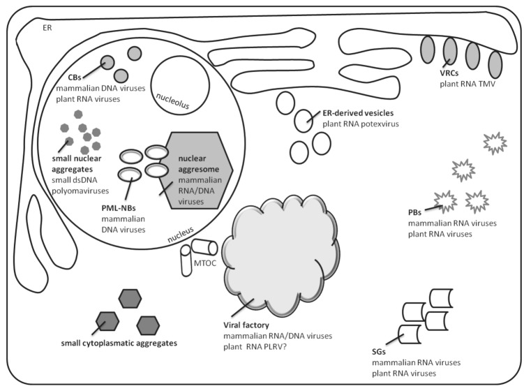
During infection, many viruses induce cellular remodeling, resulting in the formation of insoluble aggregates/inclusions, usually containing viral structural proteins. Identification of aggregates has become a useful diagnostic tool for certain viral infections. There is wide variety of viral aggregates, which differ by their location, size, content and putative function. The role of aggregation in the context of a specific virus is often poorly understood, especially in the case of plant viruses. The aggregates are utilized by viruses to house a large complex of proteins of both viral and host origin to promote virus replication, translation, intra- and intercellular transportation. Aggregated structures may protect viral functional complexes from the cellular degradation machinery. Alternatively, the activation of host defense mechanisms may involve sequestration of virus components in aggregates, followed by their neutralization as toxic for the host cell. The diversity of virus-induced aggregates in mammalian and plant cells is the subject of this review.
Figures
Similar articles
-
Host endomembrane recruitment for plant RNA virus replication.Curr Opin Virol. 2012 Dec;2(6):683-90. doi: 10.1016/j.coviro.2012.10.003. Epub 2012 Nov 2. Curr Opin Virol. 2012. PMID: 23123078 Free PMC article. Review.
-
Pathogenesis mediated by proviral host factors involved in translation and replication of plant positive-strand RNA viruses.Curr Opin Virol. 2016 Apr;17:11-18. doi: 10.1016/j.coviro.2015.11.004. Epub 2015 Nov 30. Curr Opin Virol. 2016. PMID: 26651023 Review.
-
Composition of plant virus RNA replicase complexes.Curr Opin Virol. 2012 Dec;2(6):669-75. doi: 10.1016/j.coviro.2012.09.014. Epub 2012 Oct 18. Curr Opin Virol. 2012. PMID: 23083891 Review.
-
Phytosterol metabolism in plant positive-strand RNA virus replication.Plant Cell Rep. 2022 Feb;41(2):281-291. doi: 10.1007/s00299-021-02799-x. Epub 2021 Oct 19. Plant Cell Rep. 2022. PMID: 34665312 Review.
-
A guide to viral inclusions, membrane rearrangements, factories, and viroplasm produced during virus replication.Adv Virus Res. 2007;70:101-82. doi: 10.1016/S0065-3527(07)70004-0. Adv Virus Res. 2007. PMID: 17765705 Free PMC article. Review.
Cited by
-
The P2 of Wheat yellow mosaic virus rearranges the endoplasmic reticulum and recruits other viral proteins into replication-associated inclusion bodies.Mol Plant Pathol. 2014 Jun;15(5):466-78. doi: 10.1111/mpp.12109. Epub 2014 Feb 8. Mol Plant Pathol. 2014. PMID: 24304930 Free PMC article.
-
Aggregation of Adenovirus 2 in Source Water and Impacts on Disinfection by Chlorine.Food Environ Virol. 2016 Jun;8(2):148-55. doi: 10.1007/s12560-016-9232-x. Epub 2016 Feb 24. Food Environ Virol. 2016. PMID: 26910058 Free PMC article.
-
DNA virus replication compartments.J Virol. 2014 Feb;88(3):1404-20. doi: 10.1128/JVI.02046-13. Epub 2013 Nov 20. J Virol. 2014. PMID: 24257611 Free PMC article. Review.
-
Dengue, Yellow Fever, Zika and Chikungunya epidemic arboviruses in Brazil: ultrastructural aspects.Mem Inst Oswaldo Cruz. 2021 Feb 3;115:e200278. doi: 10.1590/0074-02760200278. eCollection 2021. Mem Inst Oswaldo Cruz. 2021. PMID: 33566939 Free PMC article.
-
Human cytomegalovirus UL76 elicits novel aggresome formation via interaction with S5a of the ubiquitin proteasome system.J Virol. 2013 Nov;87(21):11562-78. doi: 10.1128/JVI.01568-13. Epub 2013 Aug 21. J Virol. 2013. PMID: 23966401 Free PMC article.
References
Publication types
MeSH terms
LinkOut - more resources
Full Text Sources
Other Literature Sources
