Review
. 2012 Oct 29;4(11):2598-635.
doi: 10.3390/v4112598.

dsRNA-dependent protein kinase PKR and its role in stress, signaling and HCV infection

Affiliations
Review

dsRNA-dependent protein kinase PKR and its role in stress, signaling and HCV infection

Stéphanie Dabo et al. Viruses. .

Abstract

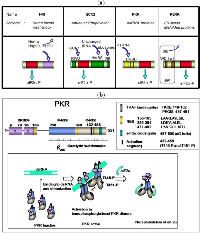
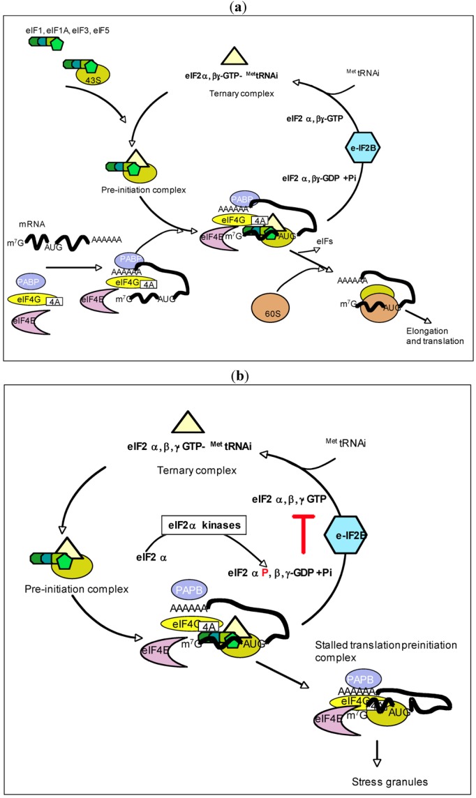
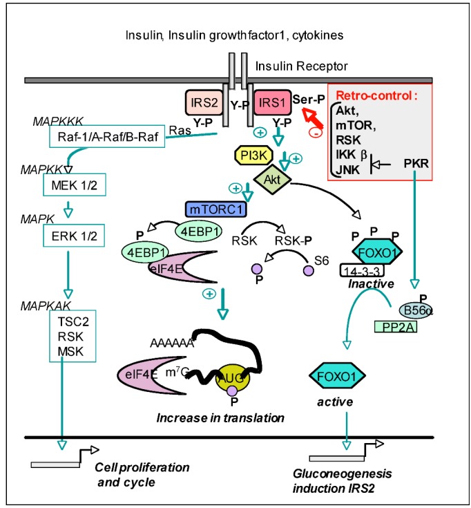
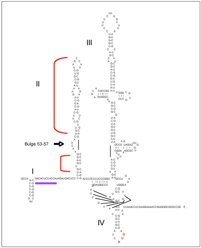
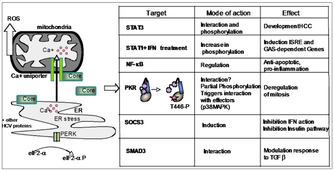
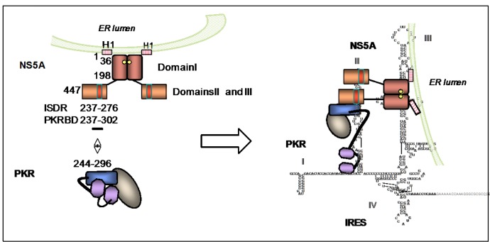
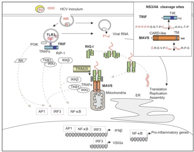
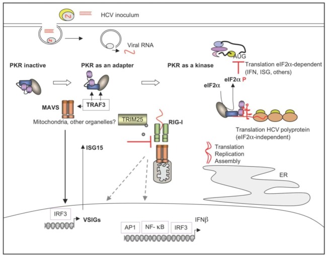
The double-stranded RNA-dependent protein kinase PKR plays multiple roles in cells, in response to different stress situations. As a member of the interferon (IFN)‑Stimulated Genes, PKR was initially recognized as an actor in the antiviral action of IFN, due to its ability to control translation, through phosphorylation, of the alpha subunit of eukaryotic initiation factor 2 (eIF2a). As such, PKR participates in the generation of stress granules, or autophagy and a number of viruses have designed strategies to inhibit its action. However, PKR deficient mice resist most viral infections, indicating that PKR may play other roles in the cell other than just acting as an antiviral agent. Indeed, PKR regulates several signaling pathways, either as an adapter protein and/or using its kinase activity. Here we review the role of PKR as an eIF2a kinase, its participation in the regulation of the NF-kB, p38MAPK and insulin pathways, and we focus on its role during infection with the hepatitis C virus (HCV). PKR binds the HCV IRES RNA, cooperates with some functions of the HCV core protein and may represent a target for NS5A or E2. Novel data points out for a role of PKR as a pro-HCV agent, both as an adapter protein and as an eIF2a-kinase, and in cooperation with the di-ubiquitin-like protein ISG15. Developing pharmaceutical inhibitors of PKR may help in resolving some viral infections as well as stress-related damages.


Figures

Figure 1
Figure 1
(a) The eIF2α-kinase family. Schematic representation of the four eIF2α-kinases: HRI, GCN2, PKR and PERK, showing their catalytic domains (in red) located at the N terminus (HRI, GCN2) or C terminus (PKR and PERK). HRI activates in response to diminution of heme levels or binding to some heat shock proteins such as Hsp90 or Hsc70. GCN2 form an inactive dimer through its region homologous to histidyl-tRNA synthetase (HisRS) and becomes activated, by change of conformation, when this domain binds uncharged tRNA, allowing its N-terminal ring finger and WD repeat domain (RWD) to bind GCN1 required for its function. It contains also a Ribosomal Binding site (RB) at its C terminus. PKR activates through two basic domains or dsRNA binding domains (DRBD) located at its N terminus. PERK is maintained in an inactive state through binding to the chaperone Bip protein (also known as Grp78 or Hsp 70-5) at its luminal N terminus (on WD repeats and Ire1-like domain). Unfolded proteins appearing in the lumen during ER stress attract Bip, which allows PERK homodimerization and activation of its cytosolic kinase domain. (b) PKR structure and mode of activation. The two 73 aa dsRNA Binding Domains (DRBD1 and DRBD2), located at the N terminus of PKR (purple) are responsible for the binding of PKR to its regulators. Mutation of the K296 residue (in subdomain II of the 253–525 catalytic domain; white bars) is sufficient to abrogate the catalytic activity of PKR. The sequences of the TRAF binding sites (grey bars) and nuclear export sequences (NES; yellow bars) is as indicated in the upper box. The eIF2α substrate binds in the helical C-lobe of PKR (blue bar) and the activation segment (black) contains the threonines 446 and 451. Lower box: Representation of the activation process of PKR upon binding to dsRNA. After binding of the first DRBD to dsRNA, binding of the second DRBD stabilizes the dsRNA/PKR complex and opens the conformation of PKR. This allows PKR dimerization through the N-lobe of its kinase domain (blue). PKR dimers activate each other by transphosphorylation of their T446 and T551 residues (red stars). The substrate (here eIF2α; green) can then access to its docking site and is positioned correctly within the catalytic domain of PKR where its acceptor site (yellow star) can receive the phosphate from ATP.
Figure 2
Figure 2
(a) eIF2 and initiation of translation. The initiation factors eIF1,eIF1A, eIF3 associate with eIF5 and the ternary complex eIF2α,β,γ-GTP-MettRNAi to form the pre‑initiation complex on the 43S ribosomal subunit. The mRNA associates with the eIF4A/4G/4A complex through its 5' monomethylated guanosine cap structure (abbreviated as m7G) and with the PolyA Binding Protein (PABP) at its polyadenylated 3'end. After association of all the components on the 43S, the preinitiation complex scans the RNA for recognition of the AUG initiation codon. This is followed by binding of MettRNAi, and hydrolysis of GTP in the ternary complex, which is thus released as the binary complex eIF2α,β,γ-GDP together with free phosphate (Pi) while the 60S ribosomal subunit binds to the RNA/43S complex. The eIFs are then released and the process of elongation and translation begins. The binary complex eIF2α,β,γ-GDP associates with the large eIF2B protein complex to be regenerated as ternary complex. It can then associate with a new MettRNAi and start a novel round of initiation of translation (adapted from [29]). (b) eIF2α kinases and control of initiation of translation. Activation of the eIF2α kinases (PKR, PERK, GCN2, HRI) under conditions of stress trigger the phosphorylation of the α subunit of eIF2 in the eIF2α,β,γ-GDP complex. This prevents the replacement of GDP by GTP within the eIF2B complex and inhibition of the initiation process. The translation pre‑initiation complex is then blocked and the different components are used for the formation of stress granules.
Figure 3
Figure 3
PKR and the Insulin signaling pathway. After binding of the insulin receptor to its different ligands, its intracytosolic chains autophosphorylate on tyrosine residues and phosphorylate the two substrates IRS1 and IRS2 on tyrosine residues to recruit the SH2‑containing Phosphoinositol-3 kinase (PI3K). Lipid products generated by PI3K bind and activate Akt, which in turn activates mTORC1. The latter phosphorylates 4EBP1 and RSK (Ribosomal S6 kinase). As a result, 4EBP1 dissociates from the cap-binding eIF4E protein and RSK phosphorylates the S6 ribosomal protein. The outcome of this is an increase in translation. Activation of the insulin receptor triggers also the Mitogen Activated Protein Kinase (MAPK) cascade (reviewed in [85]) leading to the transcription of factors required for cell proliferation and cycle. Akt also negatively regulates the Forkhead BoxO transcription factor FOXO1 by phosphorylation. This maintains FOXO1 in the cytosol in a complex with the 14-3-3 protein and prevents gluconeogenesis or transcription of IRS2 when not needed. Retro-control of the insulin pathway occurs through phosphorylation of IRS1 on serine residues by Akt, mTOR, RSK and by PKR, the latter acting through the intermediate IKKβ or JNK kinases. This prevents its interaction with the insulin receptor. However, PKR can also positively activate the insulin signaling by triggering FOXO1 dephosphorylation through the phosphorylation of B56α, the regulatory subunit of the protein phosphatase PP2A.
Figure 4
Figure 4
PKR and the HCV IRES. Secondary structure of the Internal Ribosomal Entry site (IRES), located within the first 388 nucleotides of HCV RNA. The four stem-loop domains of the IRES are numbered in Roman numerals (I, II, III and IV). The binding site for miR122 is underlined (purple). The brackets (in red), located on each side of a 53–57 bulge (arrow) indicate the protected areas from ribonuclease or 2' hydroxyl attack when PKR is present.
Figure 5
Figure 5
PKR and the HCV core protein. After translation from the HCV RNA, the HCV proteins, including the core protein, interact with the membrane of the endoplasmic reticulum (ER). Accumulation of the core protein at the ER triggers an ER stress which can lead to a PERK-mediated eIF2α phosphorylation and also a release of calcium (Ca+). The core protein binds also to the outer membrane of mitochondria where it stimulates the Calcium uniporter, thus allowing calcium uptake and generation of Reactive Oxygen Species (ROS). The core protein interacts with multiple cellular proteins and affects a number of signaling pathways. Some of its targets are shown in the left table, with indications of its mode of action and effect on the cells. The core protein interacts with PKR to trigger its partial phosphorylation (on threonine 446) which may change the conformation of PKR and favor its interaction with other partners such as p38MAPK.
Figure 6
Figure 6
PKR and the HCV NS5A protein. The 447 aa NS5A protein associates as a dimer and is anchored in plane to the cytosolic part of the ER through its amphipathic helix (H1) present in its domain I. NS5A interacts with HCV RNA through its domain I. Its domains II and III are flexible and able to interact with different proteins. The ISDR (237–276) and PKR binding domain (PKRBD: 237–302) are located in domain II (red bar). NS5A can interact with the 244–296 region of PKR corresponding to the N lobe of the catalytic domain of PKR. This interaction may benefit from the binding of NS5A and PKR to the domains III and II of HCV RNA, respectively.
Figure 7
Figure 7
HCV and induction of the innate immune response. HCV activates the innate immune response through the endosomal TLR3 via dsRNA structures (derived from apoptotic of necrotic infected cells) present in the inoculum and through the intracytosolic RNA helicase RIG-I after entry of the viral RNA into the cytosol. Activation through TLR7 is not represented here. Binding of RIG-I to viral dsRNA structures triggers a change in its conformation and its dimerization/multimerization. RIG-I becomes ubiquitined (grey circles) by the E3 ligase TRIM25, which allows its interaction with the mitochondria-bound protein MAVS [157] (MAVS is also known as Cardif [158], VISA [159] and IPS-1 [160]). MAVS recruits different adapters such as members of the TRAF family and the downstream kinases IKKβ, TBK1 and IKKε which activate the transcription factors NF-κB and IRF3, respectively. In addition, the MAVS pathway can also lead to activation of AP-1 (ATF-2/C-jun) (not represented). Upon binding to dsRNA, TLR3 recruits the intracytosolic adapter TRIF, which itself recruits members of the TRAF family, the adapter RIP-1 (Receptor-Interacting Protein 1) and the NF-κB- and IRF3‑activating kinases. In addition, the TLR3 pathway triggers induction of AP-1 through PI3K/Akt. Combination of the three transcription factors is necessary for efficient induction of IFNβ, while IRF3 alone is sufficient for induction of a series of genes, referred to as VISGs (Virus Stress Inducible Genes), which will be later on responsive to IFN. NF-κB stimulates the transcription of pro-inflammatory genes. Box: The HCV NS3/4A protease has the capacity to abrogate induction of the innate immune response through the cleavage of TRIF, close to its Toll-like/IL-1 Receptor (TIR)-domain and of MAVS, close to its transmembrane domain. The sequence of the cleavage sites is shown. CARD stands for Caspase Recognition Domain.
Figure 8
Figure 8
PKR, ISG15 as modulators of IFN induction during HCV infection. HCV triggers associations of PKR with TRAF3 and MAVS early in infection. This involves the first DRBD of PKR but leaves PKR inactive as a kinase. The PKR/MAVS association leads to the activation of IRF3 and induction of VSIGs. Among these, ISG15 prevents the TRIM25-mediated ubiquitination of RIG-I and its association with MAVS, thus inhibiting the IFN induction pathway. Later in infection, PKR is activated as a kinase, phosphorylates eIF2α, with subsequent transient inhibition of the general translation including IFN and IFN-Stimulated Genes (ISGs) while the translation process of the HCV polyprotein proceeds unabated since it is eIF2α-independent.

Similar articles

Cited by

References

    1. Meurs E., Chong K., Galabru J., Thomas S.B., Kerr I.M., Williams B.R.G., Hovanessian A.G. Molecular cloning and characterization of cDNA encoding human double-stranded RNA activated protein kinase induced by interferon. Cell. 1990;62:379–390. doi: 10.1016/0092-8674(90)90374-N. - DOI - PubMed
    1. Raven J.F., Koromilas A.E. PERK and PKR: Old kinases learn new tricks. Cell Cycle. 2008;7:1146–1150. doi: 10.4161/cc.7.9.5811. - DOI - PubMed
    1. Saunders L.R., Barber G.N. The dsRNA binding protein family: Critical roles, diverse cellular functions. FASEB J. 2003;17:961–983. - PubMed
    1. Daher A., Laraki G., Singh M., Melendez-Pena C.E., Bannwarth S., Peters A.H., Meurs E.F., Braun R.E., Patel R.C., Gatignol A. TRBP control of PACT-induced phosphorylation of protein kinase R is reversed by stress. Mol. Cell Biol. 2009;29:254–265. - PMC - PubMed
    1. Nallagatla S.R., Toroney R., Bevilacqua P.C. Regulation of innate immunity through RNA structure and the protein kinase PKR. Curr. Opin. Struct. Biol. 2011;21:119–127. doi: 10.1016/j.sbi.2010.11.003. - DOI - PMC - PubMed

Publication types