

Molecular pathogenesis of neuromyelitis optica
- PMID: 23202933
- PMCID: PMC3497307
- DOI: 10.3390/ijms131012970
Molecular pathogenesis of neuromyelitis optica
Abstract
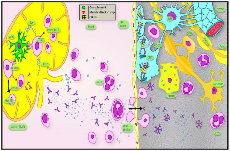
Neuromyelitis optica (NMO) is a rare autoimmune disorder, distinct from multiple sclerosis, causing inflammatory lesions in the optic nerves and spinal cord. An autoantibody (NMO IgG) against aquaporin-4 (AQP4), a water channel expressed on astrocytes is thought to be causative. Peripheral production of the antibody is triggered by an unknown process in genetically susceptible individuals. Anti-AQP4 antibody enters the central nervous system (CNS) when the blood brain barrier is made permeable and has high affinity for orthogonal array particles of AQP4. Like other autoimmune diseases, Th17 cells and their effector cytokines (such as interleukin 6) have been implicated in pathogenesis. AQP4 expressing peripheral organs are not affected by NMO IgG, but the antibody causes extensive astrocytic loss in specific regions of the CNS through complement mediated cytotoxicity. Demyelination occurs during the inflammatory process and is probably secondary to oligodendrocyte apoptosis subsequent to loss of trophic support from astrocytes. Ultimately, extensive axonal injury leads to severe disability. Despite rapid advances in the understanding of NMO pathogenesis, unanswered questions remain, particularly with regards to disease mechanisms in NMO IgG seronegative cases. Increasing knowledge of the molecular pathology is leading to improved treatment strategies.
Figures

References
-
- Allbutt T. On the opthalmoscopic signs of spinal disease. Lancet. 1870;1:76–78.
-
- Erb W. About the concurrence of optic neuritis and subacute myelitis. Arch. Psychiatr. Nervenkr. 1879;1:146–157.
-
- Devic E. Myélite subaiguë compliquée de névrite optique. Bull. Med. (Paris) 1894;8:1033–1034.
-
- Totsuka S. Clinico-pathological studies on the two cases of neuromyelitis optica (Devic’s disease) with a chronic clinical course, with especial reference on its relationship to multiple sclerosis. Folia Psychiatr. Neurol. Jpn. 1962;64:1149–1165. - PubMed
Publication types
MeSH terms
Substances
LinkOut - more resources
Full Text Sources
Other Literature Sources
