A robust procedure for distinctively visualizing zebrafish retinal cell nuclei under bright field light microscopy
- PMID: 23204114
- PMCID: PMC3636700
- DOI: 10.1369/0022155412471535
A robust procedure for distinctively visualizing zebrafish retinal cell nuclei under bright field light microscopy
Abstract
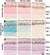
To simultaneously visualize individual cell nuclei and tissue morphologies of the zebrafish retina under bright field light microscopy, it is necessary to establish a procedure that specifically and sensitively stains the cell nuclei in thin tissue sections. This necessity arises from the high nuclear density of the retina and the highly decondensed chromatin of the cone photoreceptors, which significantly reduces their nuclear signals and makes nuclei difficult to distinguish from possible high cytoplasmic background staining. Here we optimized a procedure that integrates JB4 plastic embedding and Feulgen reaction for visualizing zebrafish retinal cell nuclei under bright field light microscopy. This method produced highly specific nuclear staining with minimal cytoplasmic background, allowing us to distinguish individual retinal nuclei despite their tight packaging. The nuclear staining is also sensitive enough to distinguish the euchromatin from heterochromatin in the zebrafish cone nuclei. In addition, this method could be combined with in situ hybridization to simultaneously visualize the cell nuclei and mRNA expression patterns. With its superb specificity and sensitivity, this method may be extended to quantify cell density and analyze global chromatin organization throughout the retina or other tissues.
Conflict of interest statement
Figures


Similar articles
-
A polychromatic staining method for epoxy embedded tissue: a new combination of methylene blue and basic fuchsine for light microscopy.Biotech Histochem. 2005 Sep-Dec;80(5-6):207-10. doi: 10.1080/10520290600560897. Biotech Histochem. 2005. PMID: 16720521
-
Embedding, serial sectioning and staining of zebrafish embryos using JB-4 resin.Nat Protoc. 2011 Jan;6(1):46-55. doi: 10.1038/nprot.2010.165. Epub 2010 Dec 16. Nat Protoc. 2011. PMID: 21212782 Free PMC article.
-
The pineal gland in wild-type and two zebrafish mutants with retinal defects.J Neurocytol. 2001 Jun;30(6):493-501. doi: 10.1023/a:1015689116620. J Neurocytol. 2001. PMID: 12037465
-
The Feulgen reaction 75 years on.Histochem Cell Biol. 1999 May;111(5):345-58. doi: 10.1007/s004180050367. Histochem Cell Biol. 1999. PMID: 10403113 Review.
-
A moving wave patterns the cone photoreceptor mosaic array in the zebrafish retina.Int J Dev Biol. 2004;48(8-9):935-45. doi: 10.1387/ijdb.041873pr. Int J Dev Biol. 2004. PMID: 15558484 Review.
Cited by
-
Photoreceptor Degeneration Accompanies Vascular Changes in a Zebrafish Model of Diabetic Retinopathy.Invest Ophthalmol Vis Sci. 2020 Feb 7;61(2):43. doi: 10.1167/iovs.61.2.43. Invest Ophthalmol Vis Sci. 2020. PMID: 32106290 Free PMC article.
-
Differential Responses of Neural Retina Progenitor Populations to Chronic Hyperglycemia.Cells. 2021 Nov 22;10(11):3265. doi: 10.3390/cells10113265. Cells. 2021. PMID: 34831487 Free PMC article.
-
Novel Animal Model of Crumbs-Dependent Progressive Retinal Degeneration That Targets Specific Cone Subtypes.Invest Ophthalmol Vis Sci. 2018 Jan 1;59(1):505-518. doi: 10.1167/iovs.17-22572. Invest Ophthalmol Vis Sci. 2018. PMID: 29368007 Free PMC article.
References
-
- Bancroft JD, Gamble M. 2008. Theory and practice of histological Techniques. 6th ed. London: Churchill Livingstone
-
- Bertero M, De Mol C. 1996. Super-resolution by data inversion. Progress in Optics. 36:129–178
-
- Bilotta J, Saszik S, Sutherland SE. 2001. Rod contributions to the electroretinogram of the dark-adapted developing zebrafish. Dev Dyn. 222:564–570 - PubMed
-
- Böcking A, Giroud F, Reith A. 1995. Consensus report of the ESACP task force on standardization of diagnostic DNA image cytometry. Anal Cell Pathol. 8:67–74 - PubMed
-
- Böhm N, Sprenger E. 1968. Fluorescence cytophotometry: a valuable method for the quantitative determination of nuclear Feulgen-DNA. Histochemie. 16:100–118 - PubMed
Publication types
MeSH terms
Substances
Grants and funding
LinkOut - more resources
Full Text Sources

