. 2013 Jan;27(1):162-71.
doi: 10.1210/me.2012-1174. Epub 2012 Nov 30.

Mitogen-inducible gene 6 triggers apoptosis and exacerbates ER stress-induced β-cell death

Affiliations

Mitogen-inducible gene 6 triggers apoptosis and exacerbates ER stress-induced β-cell death

Yi-Chun Chen et al. Mol Endocrinol. 2013 Jan.

Abstract

The increased insulin secretory burden placed on pancreatic β-cells during obesity and insulin resistance can ultimately lead to β-cell dysfunction and death and the development of type 2 diabetes. Mitogen-inducible gene 6 (Mig6) is a cellular stress-responsive protein that can negatively regulate the duration and intensity of epidermal growth factor receptor signaling and has been classically viewed as a molecular brake for proliferation. In this study, we used Mig6 heterozygous knockout mice (Mig6(+/-)) to study the role of Mig6 in regulating β-cell proliferation and survival. Surprisingly, the proliferation rate of Mig6(+/-) pancreatic islets was lower than wild-type islets despite having comparable β-cell mass and glucose tolerance. We thus speculated that Mig6 regulates cellular death. Using adenoviral vectors to overexpress or knockdown Mig6, we found that caspase 3 activation during apoptosis was dependent on the level of Mig6. Interestingly, Mig6 expression was induced during endoplasmic reticulum (ER) stress, and its protein levels were maintained throughout ER stress. Using polyribosomal profiling, we identified that Mig6 protein translation was maintained, whereas the global protein translation was inhibited during ER stress. In addition, Mig6 overexpression exacerbated ER stress-induced caspase 3 activation in vitro. In conclusion, Mig6 is transcriptionally up-regulated and resistant to global translational inhibition during stressed conditions in β-cells and mediates apoptosis in the form of caspase 3 activation. The sustained production of Mig6 protein exacerbates ER stress-induced β-cell death. Thus, preventing the induction, translation, and/or function of Mig6 is warranted for increasing β-cell survival.


Figures

Fig. 1.
Fig. 1.
Mig6+/+ and Mig6+/− mice have same similar glucose tolerance and islets morphology but different islets proliferation rates. Eight- to 10-wk-old Mig6+/+ and Mig6+/− mice (n = 6–8 per group) were submitted to (A) ip glucose tolerance testing and pancreata were assessed for (B) β-cell cross-sectional area and (C) islet morphology (insulin, red; glucagon, green; and nuclei, blue). D, Relative β-cells proliferation rates were determined in vivo by counting phospho-histone H3 (green)- and insulin (red)-stained cells normalized to total islet numbers. E, Islets proliferation rates were measured in vitro by a [3H]thymidine incorporation assay (islets from same genotype were pooled, n = 3). *, P < 0.05 between Mig6+/+ and Mig6+/−. DAPI, 4′,6-diamidino-2-phenylindole. IPGTT, IP glucose tolerance test; pH3, phospho-histone H3.
Fig. 2.
Fig. 2.
Knockdown of Mig6 attenuates etoposide-induced apoptosis, whereas overexpression of Mig6 exacerbates apoptosis. INS-1-derived 832/13 cells were transduced with adenoviruses producing either a scrambled control RNA (siCon) or a small interfering hairpin RNA sequence against Mig6 (siMig6). After 4 h of 50 nm etoposide treatment, immunoblotting was performed to determine the cleaved caspase 3 levels as an indicator of apoptosis (A and B), or cell lysates were used for a caspase 3 activity assay (C). INS-1 cells were transduced with adenoviruses carrying cmvGFP or cmvMig6. After treating with 50 nm etoposide (Etop), cell lysates were collected for immunoblotting (D and E). Data are presented as representative immunoblots and means ± sem; n = 3–4. *, P < 0.05 between etoposide-treated siCon and siMig6; #, P < 0.05 between etoposide-treated cmvGFP and cmvMig6.
Fig. 3.
Fig. 3.
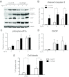
Overexpression of Mig6 exacerbates apoptosis. INS-1-derived 832/13 cells were transduced with adenoviruses carrying cmvGFP or cmvMig6. After 48 h, cells were treated with 1 μm thapsigargin (Tg) for 0, 4, or 6 h. A, Cell lysates were collected for immunoblotting to determine cleaved caspase 3 (B), phosphorylated eIF2α (C), and CHOP (D) levels. Data are presented as representative immunoblots and means ± sem; n = 3–4. *. P < 0.05 between cmvGFP and cmvMig6 groups at both 4 and 6 h of thapsigargin treatment. E, Cell death was measured by PI staining in 832/13 cells transduced with adenoviruses carrying cmvGFP or cmvMig6 and treated with DMSO or 1 μm thapsigargin for 6 h. Data represent means ± sem; n = 5. *, P < 0.05 between cmvGFP and cmvMig6 groups.
Fig. 4.
Fig. 4.
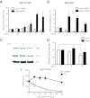
Thapsigargin-induced Mig6 mRNA expression. A, INS-1-derived 832/13 cells were treated with 1 μm thapsigargin (Tg) for the indicated times. Isolated mRNA was subjected to qRT-PCR analysis with Mig6 and Pdx-1 primers and probes. B, Isolated rat islets were treated with 1 μm thapsigargin for the indicated times and collected for qRT-PCR analysis. C and D, INS-1-derived 832/13 cells were treated with 1 μm thapsigargin for 4 and 6 h. Cell lysates were collected to determine Mig6 protein level. E, INS-1-derived 832/13 cells were pretreated with 1 μm thapsigargin for 2 h, followed by 5 μg/ml actinomycin D (ActD) treatment for 0, 20, 40, 60, 120 min, and subjected to qRT-PCR analysis. Data are presented as means ± sem; n = 3–4. *, P < 0.05 between the indicated groups.
Fig. 5.
Fig. 5.
Translation (polyribosome association) of Mig6 is maintained during ER stress. A, INS-1-derived 832/13 cells were treated with DMSO or 1 μm thapsigargin (Tg) for 6 h. Cell lysates were separated on a sucrose gradient. Ten fractions were collected while absorbance at 254 nm was continuously monitored to indicate the 40S ribosome subunits, 60S ribosome subunits, and 80S monosomes and polysomes. Samples from adjacent two fractions were pooled and subjected to RT-PCR analysis. Fractions one to four represent the monosomes, whereas fractions 5–10 represent the polysomes. Mig6 (B), ATF4 (C), and cyclinD1 (D) mRNA in the fractions were presented as percentage of unfractionated inputs. Data are presented as means ± sem; n = 4.

References

    1. Yach D, Stuckler D, Brownell KD. 2006. Epidemiologic and economic consequences of the global epidemics of obesity and diabetes. Nat Med 12:62–66 - PubMed
    1. Muoio DM, Newgard CB. 2008. Mechanisms of disease: molecular and metabolic mechanisms of insulin resistance and β-cell failure in type 2 diabetes. Nat Rev Mol Cell Biol 9:193–205 - PubMed
    1. Fonseca SG, Gromada J, Urano F. 2011. Endoplasmic reticulum stress and pancreatic β-cell death. Trends Endocrinol Metab 22:266–274 - PMC - PubMed
    1. Walter P, Ron D. 2011. The unfolded protein response: from stress pathway to homeostatic regulation. Science 334:1081–1086 - PubMed
    1. Oyadomari S, Koizumi A, Takeda K, Gotoh T, Akira S, Araki E, Mori M. 2002. Targeted disruption of the Chop gene delays endoplasmic reticulum stress-mediated diabetes. J Clin Invest 109:525–532 - PMC - PubMed

Publication types

MeSH terms

Substances