. 2013 Feb;27(2):325-35.
doi: 10.1210/me.2012-1302. Epub 2012 Nov 30.

HDAC7 inhibits osteoclastogenesis by reversing RANKL-triggered β-catenin switch

Affiliations

HDAC7 inhibits osteoclastogenesis by reversing RANKL-triggered β-catenin switch

Zixue Jin et al. Mol Endocrinol. 2013 Feb.

Abstract

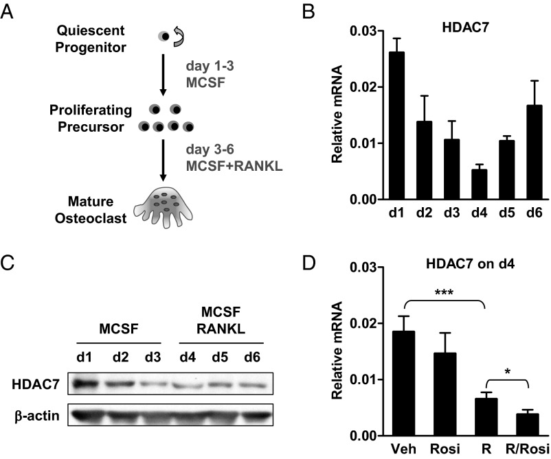
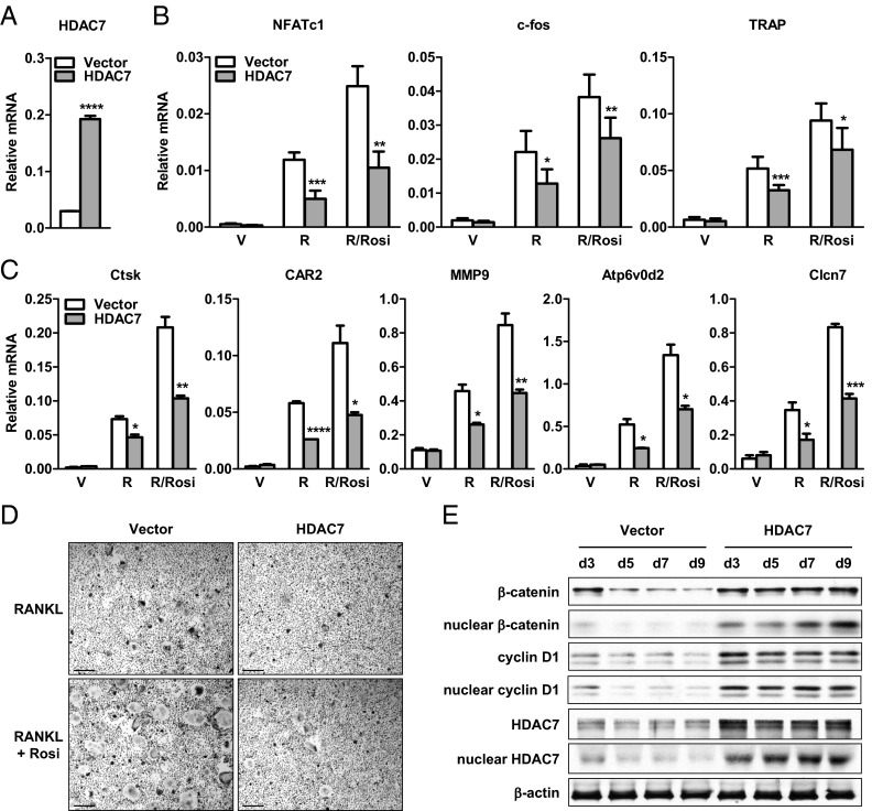
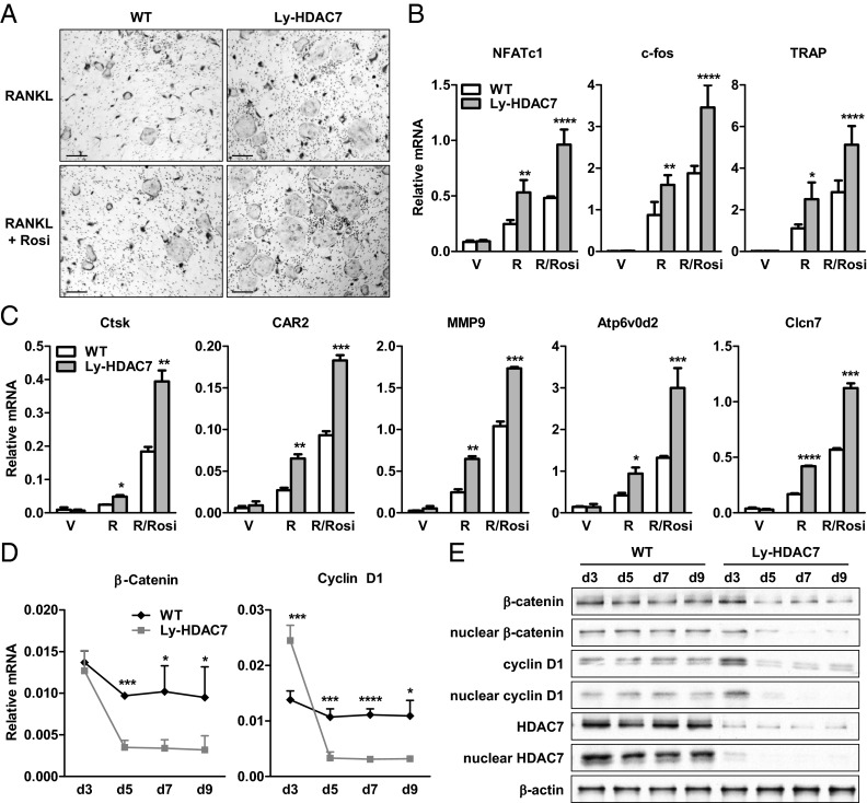
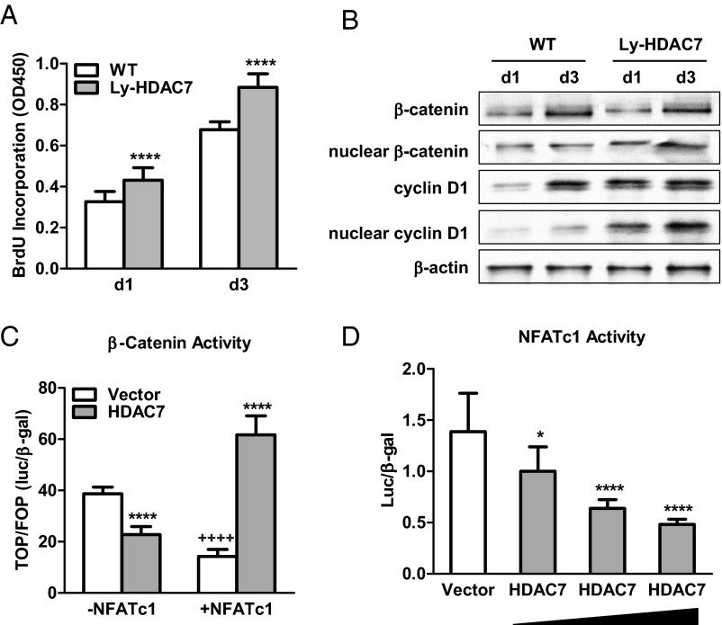
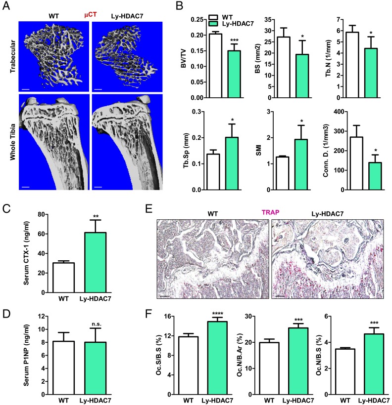
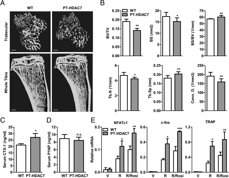
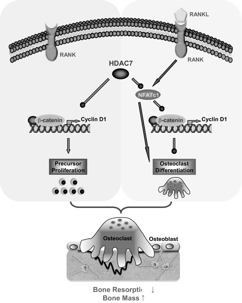
The bone-resorbing osteoclast is essential for skeletal remodeling, yet its deregulation contributes to diseases such as osteoporosis and cancer bone metastasis. Here we identify histone deacetylase 7 (HDAC7) as a key negative regulator of osteoclastogenesis and bone resorption using both in vitro cellular and molecular analyses and in vivo characterization of conditional HDAC7-knockout mice. Bone marrow osteoclast differentiation assays reveal that HDAC7 overexpression suppresses, whereas HDAC7 deletion enhances, osteoclastogenesis. Mechanistically, in the absence of receptor activator of nuclear factor κ-B ligand (RANKL), HDAC7 attenuates β-catenin function and cyclin D1 expression, thereby reducing precursor proliferation; upon RANKL activation, HDAC7 suppresses NFATc1 and prevents β-catenin down-regulation, thereby blocking osteoclast differentiation. Consequently, HDAC7 deletion in the osteoclast lineage results in a 26% reduction in bone mass (P = 0.003) owing to 102% elevated bone resorption (P = 0.01). These findings are clinically significant in light of the remarkable therapeutic potentials of HDAC inhibitors for several diseases such as cancer, diabetes, and neurodegeneration.


Figures

Fig. 1.
Fig. 1.
HDAC7 expression during osteoclast differentiation. A, A schematic diagram of the ex vivo bone marrow osteoclast differentiation assay. B, HDAC7 mRNA expression on each day of osteoclast differentiation (n = 3). C, HDAC7 protein expression on each day of osteoclast differentiation (n = 3). D, HDAC7 mRNA expression 24 h after RANKL treatment in the absence or presence of rosiglitazone (Rosi). R, RANKL; V, vehicle. Statistical analyses were performed with Student's t test and are shown as mean ± sd; *, P < 0.05; ***, P < 0.005.
Fig. 2.
Fig. 2.
HDAC7 overexpression inhibits osteoclast differentiation. Bone marrow cells from wild-type (WT) mice were transfected with HDAC7 or vector control before osteoclast differentiation with RANKL (R), with or without rosiglitazone (Rosi). A, HDAC7 mRNA expression was increased by HDAC7 transfection (n = 3). B, RANKL-induced and Rosi-stimulated expression of osteoclast transcription factors (NFATc1 and c-fos) and differentiation marker (TRAP) were attenuated by HDAC7 overexpression (n = 3). C, RANKL-induced and Rosi-stimulated expression of osteoclast resorptive activity markers were decreased by HDAC7 overexpression (n = 3). D, Osteoclast formation was suppressed by HDAC7 overexpression. Mature osteoclasts were identified as multinucleated TRAP+ (purple) cells (n = 3). Scale bar, 25μm. E, RANKL-mediated down-regulation of β-catenin and cyclin D1 proteins was abolished by HDAC7 overexpression. Whole-cell extract or nuclear extract were isolated from the differentiation cultures 3, 5, 7, or 9 d after RANKL stimulation, and immunoblotted with antibodies for β-catenin, cyclin D1, HDAC7, or β-actin. Statistical analyses were performed with Student's t test and are shown as mean ± sd; *, P < 0.05; **, P < 0.01; ***, P < 0.005; ****, P < 0.001. V, Vehicle.
Fig. 3.
Fig. 3.
HDAC7 deletion enhances osteoclast differentiation. Bone marrow cells from Ly-HDAC7 knockout mice or littermate controls were differentiated into osteoclasts with MCSF and RANKL in the absence or presence of rosiglitazone (Rosi). A, RANKL-induced and Rosi-stimulated osteoclast formation was enhanced by HDAC7 deletion. Mature osteoclasts were multinucleated TRAP+ (purple) cells (n = 3). Scale bar, 25 μm. B, RANKL-induced and Rosi-stimulated expression of osteoclast transcription factors (NFATc1 and c-fos) and differentiation marker (TRAP) were increased by HDAC7 deletion (n = 3). C, RANKL-induced and Rosi-stimulated expression of osteoclast resorptive activity markers were increased by HDAC7 deletion (n = 3). D and E, RANKL-mediated down-regulation of β-catenin and cyclin D1 mRNA (D) and protein (E) were accelerated by HDAC7 deletion (n = 3). RNA, whole-cell extract, or nuclear extract were isolated from the differentiation cultures 3, 5, 7, or 9 d after RANKL stimulation and immunoblotted with antibodies for β-catenin, cyclin D1, HDAC7, or β-actin. Statistical analyses were performed with Student's t test and are shown as mean ± sd; *, P < 0.05; **, P < 0.01; ***, P < 0.005; ****, P < 0.001. R, RANKL, V, vehicle; WT, wild type.
Fig. 4.
Fig. 4.
HDAC7 Reverses RANKL-mediated β-catenin switch by suppressing NFATc1. A, Osteoclast precursor proliferation was increased by HDAC7 deletion (n = 10). BrdU incorporation was compared between bone marrow cultures from Ly-HDAC7 knockout mice or littermate controls 1 or 3 d after MCSF treatment. B, β-Catenin and cyclin D1 protein levels during precursor proliferation were elevated by HDAC7 deletion. Whole-cell extract or nuclear extract was isolated from the bone marrow cultures 1 or 3 d after MCSF treatment, and immunoblotted with antibodies for β-catenin, cyclin D1, or β-actin. C, β-Catenin activity was inhibited by HDAC7 in the absence of NFATc1, but stimulated by HDAC7 in the presence of NFATc1 in a transient transfection assay. TOP-flash activity was normalized by FOP-flash activity control (n = 6). *, HDAC7 compared with vector control; +, −NFATc1 compared with +NFATc1. D, NFATc1 activity was suppressed in a dose-dependent manner by HDAC7 in a transient transfection assay (n = 6). Statistical analyses were performed with Student's t test and are shown as mean ± sd; *, P < 0.05; **** or ++++, P < 0.001. WT, Wild type.
Fig. 5.
Fig. 5.
Ly-HDAC7 knockout mice have low bone mass due to high bone resorption. A and B, Ly-HDAC7 mice displayed a low-bone mass phenotype. Tibias from Ly-HDAC7 mice or wild-type (WT) littermate controls (3-month-old males, n = 4) were analyzed by μCT. A, Representative images of the trabecular bone of the tibial metaphysis (top) (scale bar, 10 μm) and the entire proximal tibia (bottom) (scale bar, 1mm). B, Quantification of trabecular bone volume and architecture. Tb.N, Trabecular number; Tb.Sp, trabecular separation; SMI, structure model index; Conn.D., connectivity density. C, Serum CTX-1 was increased (3-month-old males, n = 6). D, Serum P1NP was unaltered (3-month-old males, n = 6). E and F, Bone histomorphometry analysis shows increased osteoclasts in the Ly-HDAC7 mice compared with littermate controls (3-month-old males, n =4). E, Representative images of TRAP-stained femoral sections. Osteoclasts were identified as multinucleated TRAP+ (purple) cells. Scale bar, 100 μm. F, Quantification of osteoclast surface and osteoclast number. Oc.S, osteoclast surface; B.Ar, bone area. Statistical analyses were performed with Student's t test and are shown as mean ± sd; *, P < 0.05; **, P < 0.01; ***, P < 0.005; ****, P < 0.001; n.s., nonsignificant., P > 0.05.
Fig. 6.
Fig. 6.
PT-HDAC7 knockout mice have low bone mass due to high bone resorption. A and B, PT-HDAC7 mice displayed a low-bone mass phenotype. Tibias from PT-HDAC7 mice or littermate controls (3-month-old males, n = 4) were analyzed by μCT. A, Representative images of the trabecular bone of the tibial metaphysis (top) (scale bar, 10 μm) and the entire proximal tibia (bottom) (scale bar, 1 mm). B, Quantification of trabecular bone volume and architecture. Tb.N, Trabecular number; Tb.Sp, trabecular separation; Conn.D., connectivity density. C, Serum CTX-1 was increased (3-month-old males, n = 4). D, Serum P1NP was unaltered (3-month-old males, n = 4). E, RANKL-induced and rosi-stimulated expression of osteoclast transcription factors (NFATc1 and c-fos) and functional gene (TRAP) were increased in PT-HDAC7 bone marrow differentiation culture (n = 3). R, RANKL; Rosi, rosiglitazone; V, vehicle. Statistical analyses were performed with Student's t test and are shown as mean ± sd; *, P < 0.05; **, P < 0.01; ***, P < 0.005; n.s., nonsignificant., P > 0.05.
Fig. 7.
Fig. 7.
A simplified model for how HDAC7 suppresses osteoclastogenesis. In the absence of RANKL, HDAC7 inhibits osteoclast precursor proliferation by attenuating β-catenin function and cyclin D1 expression. Upon RANKL stimulation, HDAC7 inhibits osteoclast differentiation by suppressing NFATc1 and prevents RANKL/NFATc1-mediated down-regulation of β-catenin and cyclin D1. Consequently, HDAC7 decreases osteoclastogenesis and bone resorption, thereby increasing bone mass.

Similar articles

Cited by

References

    1. Bruzzaniti A, Baron R. 2006. Molecular regulation of osteoclast activity. Rev Endocr Metab Disord 7:123–139 - PubMed
    1. Novack DV, Teitelbaum SL. 2008. The osteoclast: friend or foe? Annu Rev Pathol 3:457–484 - PubMed
    1. Wan Y. 2010. PPARγ in bone homeostasis. Trends Endocrinol Metab 21:722–728 - PubMed
    1. Grigoriadis AE, Wang ZQ, Cecchini MG, Hofstetter W, Felix R, Fleisch HA, Wagner EF. 1994. c-Fos: a key regulator of osteoclast-macrophage lineage determination and bone remodeling. Science 266:443–448 - PubMed
    1. Takayanagi H, Kim S, Koga T, Nishina H, Isshiki M, Yoshida H, Saiura A, Isobe M, Yokochi T, Inoue J, Wagner EF, Mak TW, Kodama T, Taniguchi T. 2002. Induction and activation of the transcription factor NFATc1 (NFAT2) integrate RANKL signaling in terminal differentiation of osteoclasts. Dev Cell 3:889–901 - PubMed

Publication types

MeSH terms