. 2013 Feb 1;288(5):3097-111.
doi: 10.1074/jbc.M112.362954. Epub 2012 Nov 30.

Thrombin-cleaved fragments of osteopontin are overexpressed in malignant glial tumors and provide a molecular niche with survival advantage

Affiliations

Thrombin-cleaved fragments of osteopontin are overexpressed in malignant glial tumors and provide a molecular niche with survival advantage

Yasuto Yamaguchi et al. J Biol Chem. .

Abstract

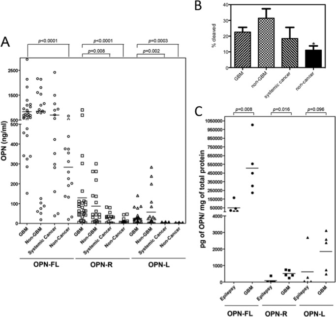
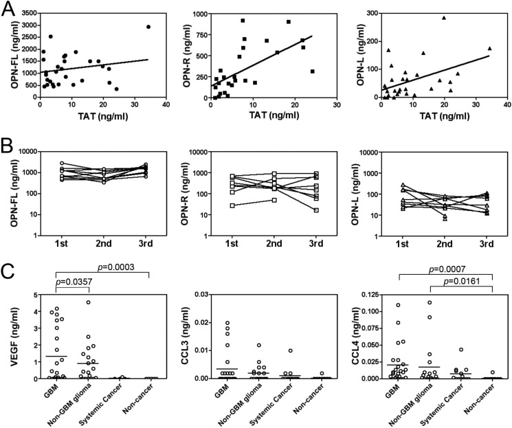
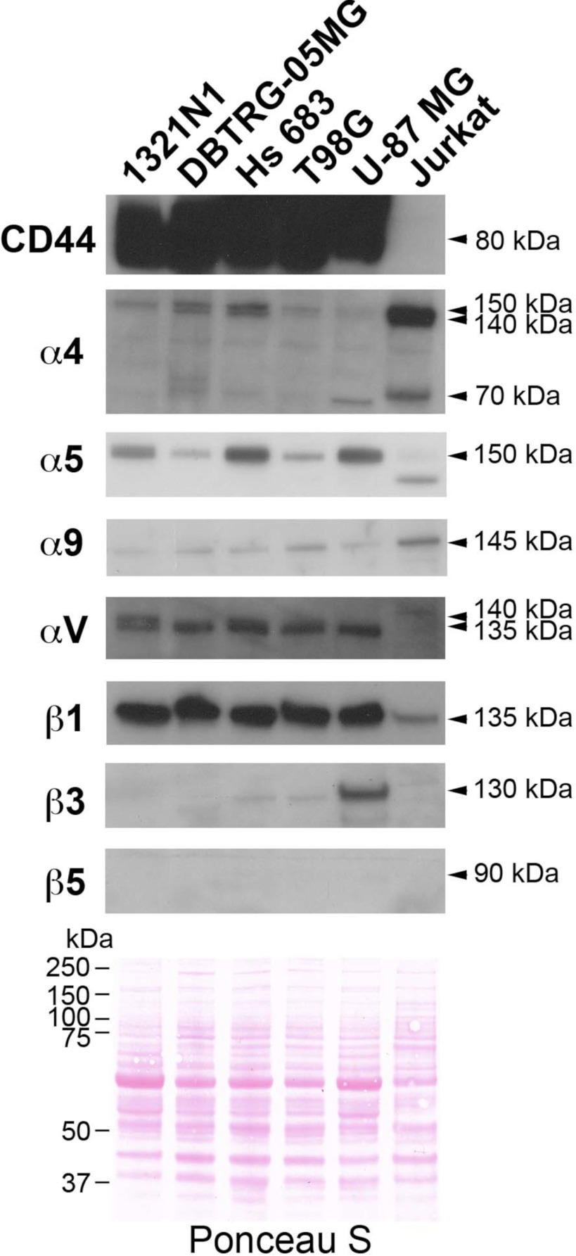
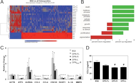
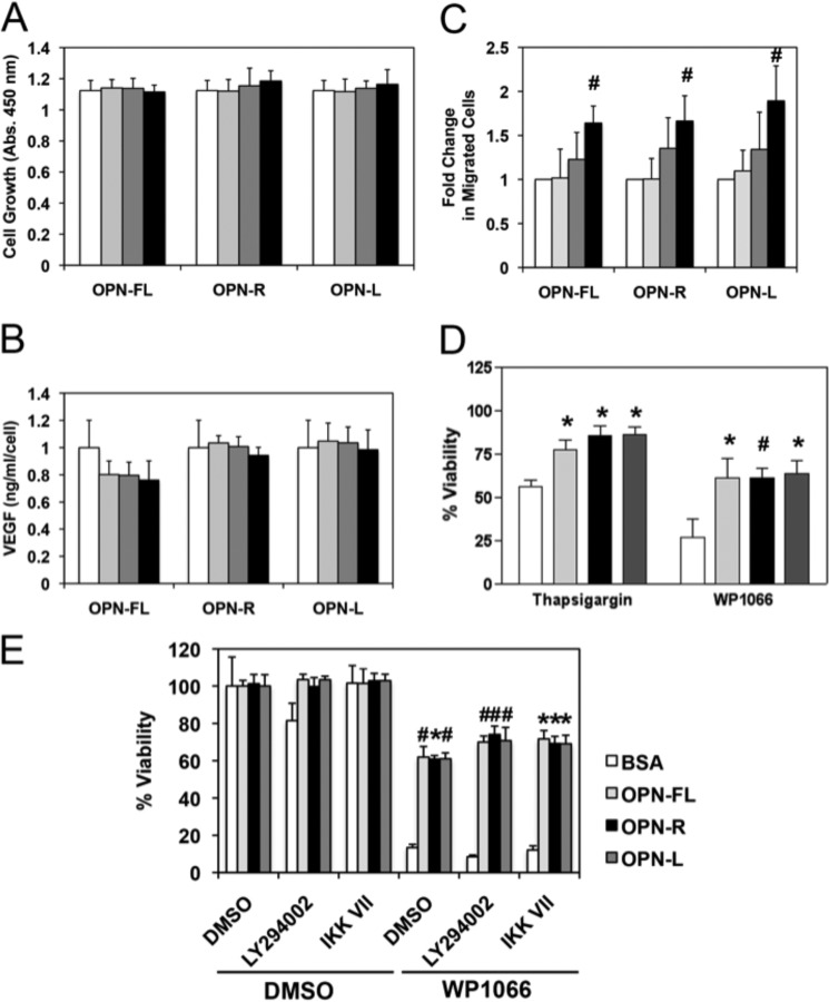
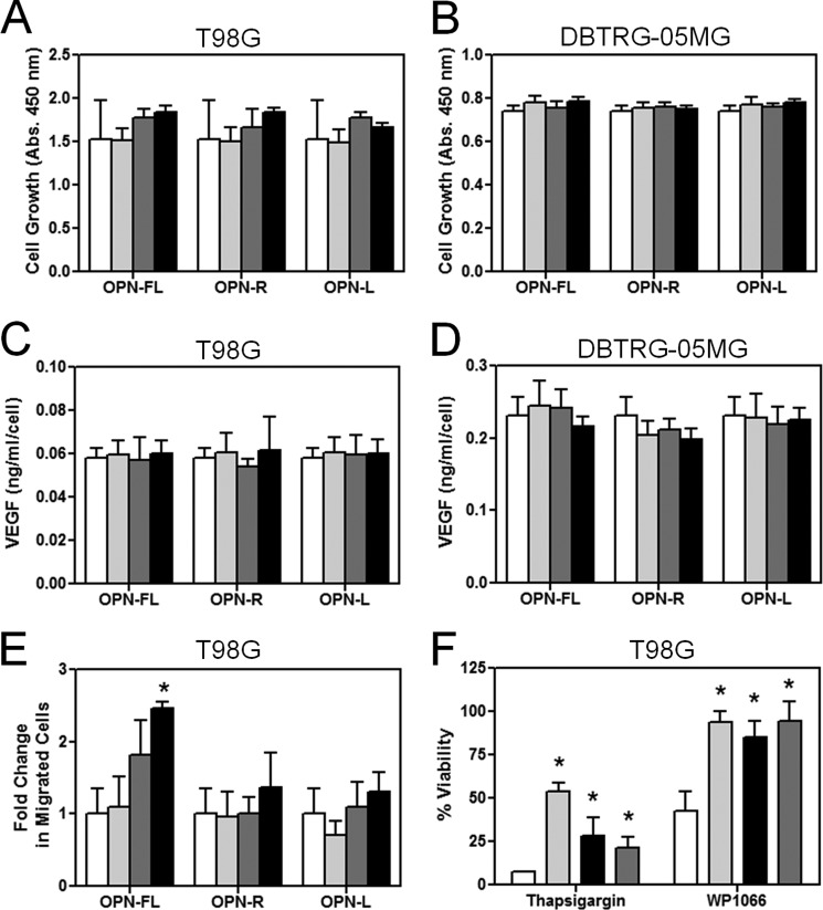
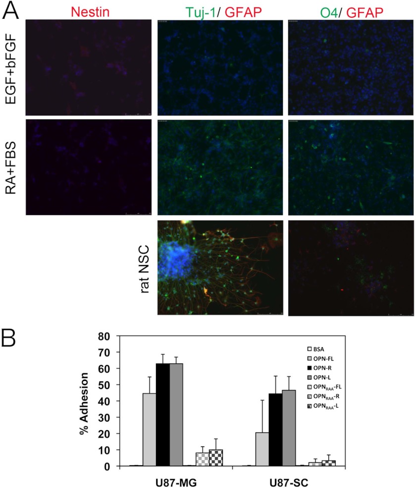
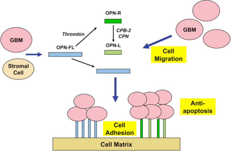
Osteopontin (OPN), which is highly expressed in malignant glioblastoma (GBM), possesses inflammatory activity modulated by proteolytic cleavage by thrombin and plasma carboxypeptidase B2 (CPB2) at a highly conserved cleavage site. Full-length OPN (OPN-FL) was elevated in cerebrospinal fluid (CSF) samples from all cancer patients compared with noncancer patients. However, thrombin-cleaved OPN (OPN-R) and thrombin/CPB2-double-cleaved OPN (OPN-L) levels were markedly increased in GBM and non-GBM gliomas compared with systemic cancer and noncancer patients. Cleaved OPN constituted ∼23 and ∼31% of the total OPN in the GBM and non-GBM CSF samples, respectively. OPN-R was also elevated in GBM tissues. Thrombin-antithrombin levels were highly correlated with cleaved OPN, but not OPN-FL, suggesting that the cleaved OPN fragments resulted from increased thrombin and CPB2 in this extracellular compartment. Levels of VEGF and CCL4 were increased in CSF of GBM and correlated with the levels of cleaved OPN. GBM cell lines were more adherent to OPN-R and OPN-L than OPN-FL. Adhesion to OPN altered gene expression, in particular genes involved with cellular processes, cell cycle regulation, death, and inflammation. OPN and its cleaved forms promoted motility of U-87 MG cells and conferred resistance to apoptosis. Although functional mutation of the RGD motif in OPN largely abolished these functions, OPN(RAA)-R regained significant cell binding and signaling function, suggesting that the SVVYGLR motif in OPN-R may substitute for the RGD motif if the latter becomes inaccessible. OPN cleavage contributes to GBM development by allowing more cells to bind in niches where they acquire anti-apoptotic properties.


Figures

FIGURE 1.
FIGURE 1.
Thrombin-cleaved fragments of OPN significantly increased in both GBM CSF and tissue. A, CSF samples from GBM patients (n = 29) were compared with CSF samples from non-GBM (n = 20), systemic cancer (n = 14), and noncancer patients (n = 15) for levels of OPN-FL (○), OPN-R (□), and OPN-L (▵). Bars represent the mean value of each group. Each point represents CSF from an individual patient. B, fraction of cleaved OPN was significantly increased in GBM and epilepsy samples compared with noncancer samples. Data were calculated from the results shown in A and shown as mean ± S.E. *, p < 0.05. C, levels of OPN-FL (●), OPN-R (■), and OPN-L (▴) in epileptic and GBM tissues determined by ELISA (n = 5 each).
FIGURE 2.
FIGURE 2.
Thrombin-cleaved OPN in CSF from GBM patients correlates with TAT levels as well as VEGF, CCL3, and CCL4. A, CSF from an independent cohort of 30 GBM patients had the levels of OPN isoforms and TAT determined. Left panel, OPN-FL (●); center panel, OPN-R (■); right panel, OPN-L (▴). B, OPN-R (center panel) and OPN-L (right panel) levels in CSF from GBM patients vary more than OPN-FL levels (left panel). CSF was obtained from 10 GBM patients on three separate occasions at least 1 month apart, and the levels of OPN-FL, OPN-R, and OPN-L were determined. The x axis is marked with the occasion when the samples were acquired. C, VEGF and CCL4 levels were increased in GBM. Levels of VEGF (left panel), CCL3 (center panel), and CCL4 (right panel) in CSF from GBM (n = 24), non-GBM (n = 18), systemic cancer (n = 13), and noncancer patients (n = 9) were determined by ELISA. Bars represent the mean value of each group.
FIGURE 3.
FIGURE 3.
Expression pattern of integrins and CD44 in human glioma cells and Jurkat cells. Different glioma cells showed different patterns of integrin expression but all could be readily distinguished from Jurkat cells. Cell lysates were separated by SDS-PAGE before detection of adhesion molecules by Western blotting. Equal loading was demonstrated by visualizing the proteins with Ponceau S staining (bottom panel).
FIGURE 4.
FIGURE 4.
Adherence of human glioma cells to OPN-R and OPN-L compared with OPN-FL. Carboxyfluorescein diacetate succinimidyl ester-labeled cells were tested for their ability to bind to different forms of OPN. The data represent the mean ± S.D. of at least three independent experiments. #, p < 0.05 compared with OPN-FL; *, p < 0.05 compared with OPNRAA-FL by paired Student's t test.
FIGURE 5.
FIGURE 5.
OPN binding altered gene expression in U-87 MG cells. A, heat map showing clustering of genes changed by >1.5-fold with p < 0.005 when U-87 MG cells bound to BSA were compared with U-87 MG cells bound to all OPNs determined by ANOVA. Both samples (y axis) and genes (x axis) were allowed to cluster unsupervised. The color scale of the log2 ratios is shown at the bottom. B, Gene Ontology classification. C, mRNA for nine genes shown were checked by quantitative PCR to validate the data from the microarray. The data were normalized to the levels of GAPDH and expressed as mean ± S.D. of relative gene expression to BSA. #, p < 0.05; *, p < 0.01 compared with BSA by paired Student's t test. D, determination of CCL3 protein level in 2 day-conditioned medium from U-87-MG cells bound to BSA and OPN by ELISA. #, p < 0.05 compared with BSA by paired Student's t test.
FIGURE 6.
FIGURE 6.
OPN promoted U-87 MG cell migration and conferred resistance to cell apoptosis. U-87 MG cells were treated with different concentrations of OPN-FL, OPN-R, and OPN-L (0 nm, white; 1 nm, light gray; 10 nm, dark gray; and 100 nm, black) for 2 days. A, cell proliferation was determined by CKK-8 assay. B, VEGF production was determined by assaying VEGF concentration in the conditioned media by ELISA, and the overall value normalized by the cell number was determined in A. C, dose-dependent enhancement of U-87 MG cell motility by OPN using the transwell chemotaxis assay. D, U-87 MG cells adhered to BSA (white), OPN-FL (light gray), OPN-R (black), and OPN-L (dark gray) are protected against apoptosis induced by 10 μm thapsigargin or 30 μm WP-1066. E, U-87 MG cells adhered to BSA (white), OPN-FL (light gray), OPN-R (black), and OPN-L (dark gray) are protected against apoptosis induced by 30 μm WP1066, even in the presence of 10 μm LY294002 or 1 μm IKK VII, inhibitors of PI3K and IKK, respectively. Their overall viability was calculated by comparison with data from untreated cells. Data are shown as mean ± S.D. and calculated from at least three independent experiments. *, p < 0.05; #, p < 0.01 compared with BSA by one-way ANOVA with post hoc Dunnett's.
FIGURE 7.
FIGURE 7.
OPN promoted T98G cell migration and conferred resistance to cell apoptosis in T98G cells. T98G and DBTRG-05MG cells were treated with different concentrations of OPN-FL, OPN-R, and OPN-L (0 nm, white; 1 nm, light gray; 10 nm, dark gray; and 100 nm, black) for 2 days. A, T98G. B, DBTRG-05MG. Cell proliferation was determined by CKK-8 assay. C, T98G. D, DBTRG-05MG. VEGF production was determined by assaying VEGF concentration in the conditioned media by ELISA, and the overall value was normalized by the cell number determined in A or B. E, dose-dependent enhancement of T98G cell motility by OPN-FL using the transwell chemotaxis assay. F, T98G cells adhered to BSA (white), OPN-FL (light gray), OPN-R (black), and OPN-L (dark gray) are protected against apoptosis induced by 15 μm thapsigargin or 10 μm WP-1066. Their overall viability was calculated by comparison with data from untreated cells. Data are shown as mean ± S.D. *, p < 0.05, compared with BSA by one-way ANOVA with post hoc Dunnett's.
FIGURE 8.
FIGURE 8.
U-87 SC cells were differentiated into Tuj-1-positive neuron and O4-positive oligodendrocyte and bound to OPN-R and OPN-L more than OPN-FL. A, characterization of U-87 SC cells in expansion (20 ng/ml EGF and basic FGF, upper panels) and differentiation conditions (1 μm retinoic acid and 1% FBS, middle panels). The cells were stained for neural stem cell (Nestin), neuron (Tuj-1), oligodendrocyte (O4), and astrocyte (GFAP) markers. The individual cells were visualized with Hoechst 33342. Control of rat neural stem cells under the same differentiation conditions is shown (bottom panels). B, adherence of U-87 MG and U-87 SC cells to OPN-R and OPN-L compared with OPN-FL. Data are shown as mean ± S.D. and calculated from at least three independent experiments.
FIGURE 9.
FIGURE 9.
Model depicting the role of OPN isoforms in GBM. Both GBM and stromal cells secrete OPN, which is cleaved by thrombin or double cleaved by thrombin and CPB2 or CPN to generate OPN-R and OPN-L. All three OPN isoforms cause cell migration, increased cell adhesion, and inhibit apoptosis, thereby creating a niche that confers survival advantages on the GBM cells.

Similar articles

Cited by

References

    1. Denhardt D. T., Noda M., O'Regan A. W., Pavlin D., Berman J. S. (2001) Osteopontin as a means to cope with environmental insults. Regulation of inflammation, tissue remodeling, and cell survival. J. Clin. Invest. 107, 1055–1061 - PMC - PubMed
    1. Han M. H., Hwang S. I., Roy D. B., Lundgren D. H., Price J. V., Ousman S. S., Fernald G. H., Gerlitz B., Robinson W. H., Baranzini S. E., Grinnell B. W., Raine C. S., Sobel R. A., Han D. K., Steinman L. (2008) Proteomic analysis of active multiple sclerosis lesions reveals therapeutic targets. Nature 451, 1076–1081 - PubMed
    1. Wang K. X., Denhardt D. T. (2008) Osteopontin. Role in immune regulation and stress responses. Cytokine Growth Factor Rev. 19, 333–345 - PubMed
    1. El-Tanani M. K., Campbell F. C., Kurisetty V., Jin D., McCann M., Rudland P. S. (2006) The regulation and role of osteopontin in malignant transformation and cancer. Cytokine Growth Factor Rev. 17, 463–474 - PubMed
    1. Minn A. J., Gupta G. P., Siegel P. M., Bos P. D., Shu W., Giri D. D., Viale A., Olshen A. B., Gerald W. L., Massagué J. (2005) Genes that mediate breast cancer metastasis to lung. Nature 436, 518–524 - PMC - PubMed

Publication types

MeSH terms