. 2013 Jan 11;288(2):1032-46.
doi: 10.1074/jbc.M112.426957. Epub 2012 Nov 30.

Molecular mechanisms underlying cardiac protein phosphatase 2A regulation in heart

Affiliations

Molecular mechanisms underlying cardiac protein phosphatase 2A regulation in heart

Sean T DeGrande et al. J Biol Chem. .

Abstract

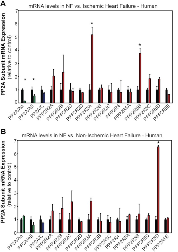
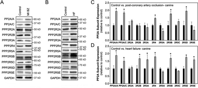
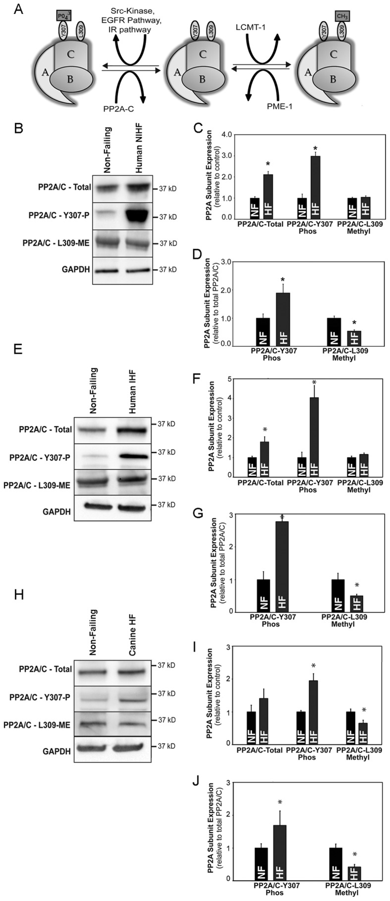
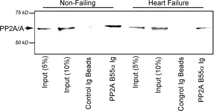
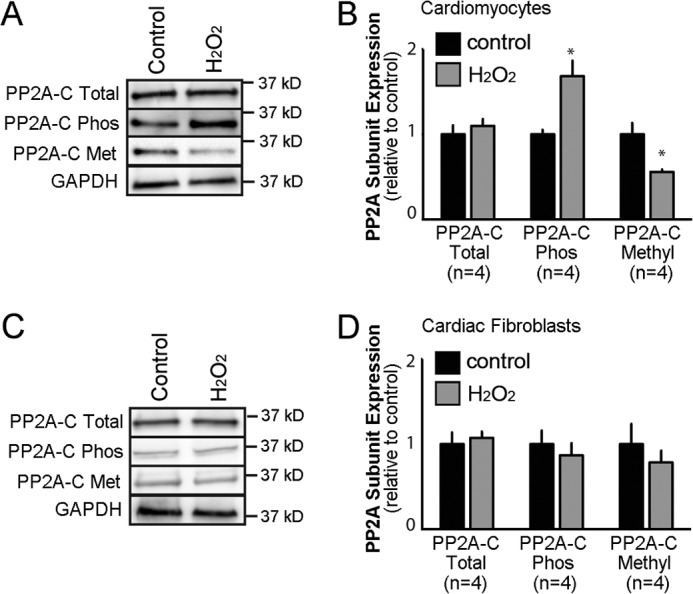
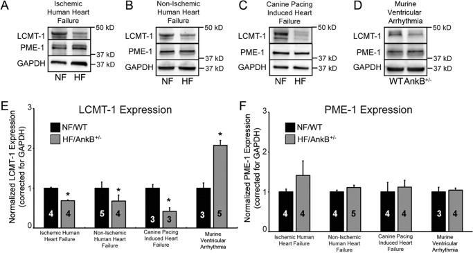
Kinase/phosphatase balance governs cardiac excitability in health and disease. Although detailed mechanisms for cardiac kinase regulation are established, far less is known regarding cardiac protein phosphatase 2A (PP2A) regulation. This is largely due to the complexity of the PP2A holoenzyme structure (combinatorial assembly of three subunit enzyme from >17 subunit genes) and the inability to segregate "global" PP2A function from the activities of multiple "local" holoenzyme populations. Here we report that PP2A catalytic, regulatory, and scaffolding subunits are tightly regulated at transcriptional, translational, and post-translational levels to tune myocyte function at base line and in disease. We show that past global read-outs of cellular PP2A activity more appropriately represent the collective activity of numerous individual PP2A holoenzymes, each displaying a specific subcellular localization (dictated by select PP2A regulatory subunits) as well as local specific post-translational catalytic subunit methylation and phosphorylation events that regulate local and rapid holoenzyme assembly/disassembly (via leucine carboxymethyltransferase 1/phosphatase methylesterase 1 (LCMT-1/PME-1). We report that PP2A subunits are selectively regulated between human and animal models, across cardiac chambers, and even within specific cardiac cell types. Moreover, this regulation can be rapidly tuned in response to cellular activation. Finally, we report that global PP2A is altered in human and experimental models of heart disease, yet each pathology displays its own distinct molecular signature though specific PP2A subunit modulatory events. These new data provide an initial view into the signaling pathways that govern PP2A function in heart but also establish the first step in defining specific PP2A regulatory targets in health and disease.


Figures

FIGURE 1.
FIGURE 1.
Defining PP2A family subunits in human heart. A, PP2A holoenzyme is shown. The model of PP2A holoenzyme consists of a scaffolding, catalytic, and regulatory subunit. B, human PP2A isoform descriptions include subunit type, gene name, related names, and PP2A subfamily. C, cardiac PP2A subunit transcripts are shown. Primers designed to amplify the specific subunit noted above were used to determine the presence of PP2A subunit transcripts in non-failing human heart. PCR products are shown, and a plus sign indicates the detection of a PCR product.
FIGURE 2.
FIGURE 2.
Differential expression of PP2A subunits in human heart chambers and animal species. A and B, shown is PP2A subunit expression in human heart chambers. Expression of PP2A subunits across human heart chambers is shown. For panel A, * denotes p < 0.05 for each subunit relative to expression in human LV (n = 3). C and D, shown is PP2A subunit expression across species. Expression of PP2A subunits in healthy human, canine, rat, and mouse LV is shown. In D, * denotes p < 0.05 for each subunit relative to human LV (n = 3). In A–D, band densities were normalized to GAPDH, and data are shown relative to expression levels in non-failing human LV.
FIGURE 3.
FIGURE 3.
Differential subcellular localization of PP2A regulatory subunits in heart. Isolated adult mouse cardiomyocytes were analyzed by confocal microscopy for distribution of PP2A subunits. A, PP2A/A. B, PP2A/C. C, PPP2R3A is in red; the asterisk denotes the site of the nucleus, arrows indicate Z-line, and arrowheads indicate M-line. D, PPP2R3A is in red, and α-actinin is in blue. E, PPP2R4 is in (green). F, PPP2R4 is in green, and α-actinin is in red. G, PPP2R5C is in red. H, PPP2R5C is in red, and α-actinin is in green. I, PPP2R5E is in blue. J, PPP2R5E is in blue, and α-actinin is in purple. Each experiment was done in triplicate using myocytes isolated from three different WT mice. Bar = 10 μm.
FIGURE 4.
FIGURE 4.
Differential PP2A subunit regulation in ischemic and non-ischemic heart disease. Shown is PP2A subunit expression in non-failing (NF) human LV and in LV of human hearts in end stage ischemic heart failure (IHF) (A) or in end stage non-ischemic heart failure (nIHF) (B). C and D, densitometry analysis describes PP2A subunit expression levels in non-failing human LV and in the LV of human hearts in end stage ischemic heart failure (IHF) (C) or in end stage non-ischemic heart failure (nIHF) (D). In all experiments, GAPDH was utilized as a loading control. n = 5 for all experiments, and the asterisk denotes p < 0.05 compared with human LV.
FIGURE 5.
FIGURE 5.
Transcriptional regulation of PP2A subunits in human heart failure. A, shown are PP2A isoform mRNA levels in LV samples from human non-failing (black bars) and ischemic heart failure. B, PP2A isoform mRNA levels in LV samples from human non-failing (black bars) and non-ischemic heart failure are shown. n = 3 for all experiments, and an asterisk denotes p < 0.05 compared with human non-failing sample. Values were normalized to GAPDH as an internal amplification control and are expressed as levels compared with non-failing samples.
FIGURE 6.
FIGURE 6.
Differential PP2A subunit regulation in canine cardiovascular disease. Shown is PP2A subunit expression in canine LV. Experiments in panels A and C represent data from control versus border zone (BZ) tissue 5 days post coronary artery occlusion (n = 5, p < 0.05). Experiments in panels B and D represent data from non-failing versus heart failure. In all experiments, GAPDH was used as a loading control (n = 5, p < 0.05). MI, myocardial infarction.
FIGURE 7.
FIGURE 7.
Post-translational regulation of PP2A subunits at base line and in disease. A, a model of post-translational modifications of the PP2A catalytic subunit is shown. Tyr-307 phosphorylation of PP2A/C results in generalized inhibition of phosphatase activity; Leu-309 methylation (regulated by LCMT-1 and PME-1) results in the enhanced recruitment of the PPP2R2A and PPP2R2B into the holoenzyme. Shown are expression levels of total, phosphorylated (Thr-307), and methylated (Leu-309) PP2A catalytic subunit in LV samples of non-failing (NF) and non-ischemic human heart failure (B and C; n = 5; p < 0.05), non-failing and ischemic human heart failure (E and F, n = 5 non-failing, n = 4 ischemic HF, p < 0.05), and canine control and heart failure samples (H–I, n = 4/group, p < 0.05). D, G, and J, adjusted phosphorylated and methylated catalytic subunit activities based on total PP2A catalytic subunit expression in non-failing and heart failure samples from human and canine heart are shown. GAPDH was used as an internal loading control. IR, insulin response.
FIGURE 8.
FIGURE 8.
Decreased assembly of PP2A enzyme with B55 regulatory subunit in heart failure. Shown are co-immunoprecipitation experiments from non-failing and failing canine LV tissue lysate using B55α-specific Ig (lanes 1–2, and 5 and 6 represent 5 and 10% of experimental input). Note that PP2A B55α Ig immunoprecipitated reduced PP2A/A subunit in failing canine tissue compared with non-failing tissue even though PP2A/A and PP2A/C levels were elevated in the canine heart failure model. Molecular weight markers are shown in the figure. Identical data were observed in multiple biochemical experiments.
FIGURE 9.
FIGURE 9.
Cardiac cell type-specific regulation of PP2A post-translational regulation. A, shown is expression of the PP2A catalytic subunit in purified populations of primary ventricular cardiomyocytes ± treatment with H2O2 to induce ROS production. Immunoblots also depict relative expression of PP2A catalytic subunit phosphorylation and methylation ±H2O2. GAPDH was utilized as internal loading control. B, shown are mean expression levels of the catalytic subunit and modified forms ± H2O2 from purified primary cardiomyocytes (n = 4; * represents p < 0.05 for each antibody ± H2O2). C, shown is expression of PP2A catalytic subunit in purified populations of primary cardiac fibroblasts ± H2O2. Immunoblots also depict relative expression of PP2A catalytic subunit phosphorylation and methylation ± H2O2. GAPDH was utilized as internal loading control. D, mean expression levels are shown of the catalytic subunit and modified forms ± H2O2 from purified primary fibroblasts (n = 4; p = N.S. for each group ± H2O2).
FIGURE 10.
FIGURE 10.
Decreased catalytic subunit methylation is linked with decreased LCMT-1 expression. A–D, shown are representative immunoblots of LCMT-1 and PME-1 expression in whole heart lysates from ischemic heart failure, non-ischemic human heart failure, canine pacing-induced heart failure, and a mouse model of a human inheritable ventricular arrhythmia syndrome. NF, non-failing. E and F, shown is densitometry analysis indicating the expression level of LCMT-1 and PME-1 expression in ischemic heart failure, non-ischemic human heart failure, canine pacing-induced heart failure, and a mouse model of a human inheritable ventricular arrhythmia syndrome relative to wild type. In all experiments, GAPDH was used as a loading control. N values are noted in the figure, and p < 0.05 was considered statistically significant.

References

    1. He B. J., Joiner M. L., Singh M. V., Luczak E. D., Swaminathan P. D., Koval O. M., Kutschke W., Allamargot C., Yang J., Guan X., Zimmerman K., Grumbach I. M., Weiss R. M., Spitz D. R., Sigmund C. D., Blankesteijn W. M., Heymans S., Mohler P. J., Anderson M. E. (2011) Oxidation of CaMKII determines the cardiotoxic effects of aldosterone. Nat. Med. 17, 1610–1618 - PMC - PubMed
    1. Chelu M. G., Sarma S., Sood S., Wang S., van Oort R. J., Skapura D. G., Li N., Santonastasi M., Müller F. U., Schmitz W., Schotten U., Anderson M. E., Valderrábano M., Dobrev D., Wehrens X. H. (2009) Calmodulin kinase II-mediated sarcoplasmic reticulum Ca2+ leak promotes atrial fibrillation in mice. J. Clin. Invest. 119, 1940–1951 - PMC - PubMed
    1. Swaminathan P. D., Purohit A., Soni S., Voigt N., Singh M. V., Glukhov A. V., Gao Z., He B. J., Luczak E. D., Joiner M. L., Kutschke W., Yang J., Donahue J. K., Weiss R. M., Grumbach I. M., Ogawa M., Chen P. S., Efimov I., Dobrev D., Mohler P. J., Hund T. J., Anderson M. E. (2011) Oxidized CaMKII causes cardiac sinus node dysfunction in mice. J. Clin. Invest. 121, 3277–3288 - PMC - PubMed
    1. Bers D. M. (2011) Ca2+-calmodulin-dependent protein kinase II regulation of cardiac excitation-transcription coupling. Heart Rhythm 8, 1101–1104 - PMC - PubMed
    1. Reiken S., Gaburjakova M., Gaburjakova J., He Kl K. L., Prieto A., Becker E., Yi Gh G. H., Wang J., Burkhoff D., Marks A. R. (2001) β-Adrenergic receptor blockers restore cardiac calcium release channel (ryanodine receptor) structure and function in heart failure. Circulation 104, 2843–2848 - PubMed

Publication types