. 2013 Jan 18;288(3):1871-82.
doi: 10.1074/jbc.M112.396200. Epub 2012 Nov 30.

Arabidopsis calmodulin-binding protein IQ67-domain 1 localizes to microtubules and interacts with kinesin light chain-related protein-1

Affiliations

Arabidopsis calmodulin-binding protein IQ67-domain 1 localizes to microtubules and interacts with kinesin light chain-related protein-1

Katharina Bürstenbinder et al. J Biol Chem. .

Abstract

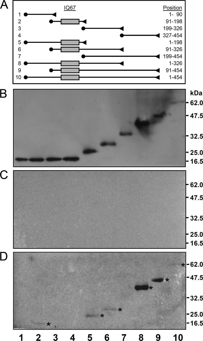
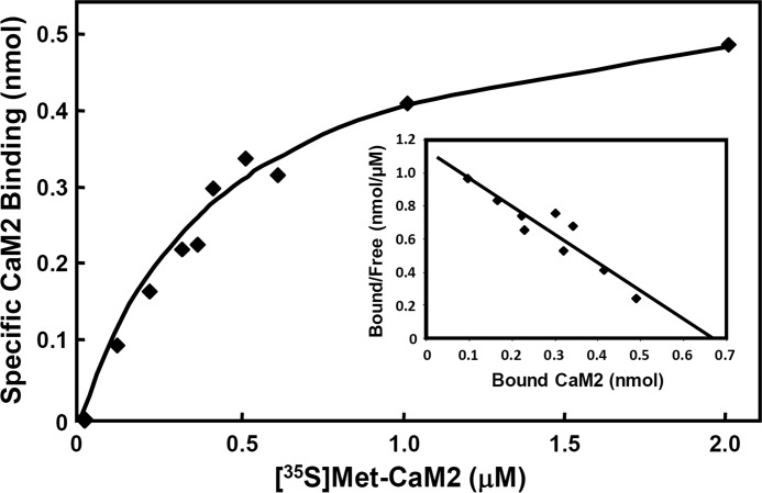
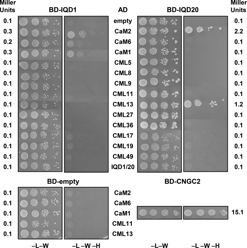
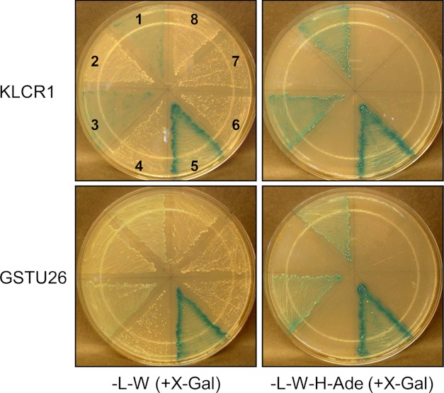
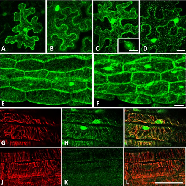
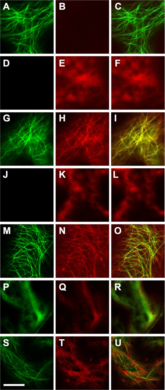
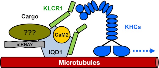
Calcium (Ca(2+)) is a key second messenger in eukaryotes and regulates diverse cellular processes, most notably via calmodulin (CaM). In Arabidopsis thaliana, IQD1 (IQ67 domain 1) is the founding member of the IQD family of putative CaM targets. The 33 predicted IQD proteins share a conserved domain of 67 amino acids that is characterized by a unique arrangement of multiple CaM recruitment motifs, including so-called IQ motifs. Whereas IQD1 has been implicated in the regulation of defense metabolism, the biochemical functions of IQD proteins remain to be elucidated. In this study we show that IQD1 binds to multiple Arabidopsis CaM and CaM-like (CML) proteins in vitro and in yeast two-hybrid interaction assays. CaM overlay assays revealed moderate affinity of IQD1 to CaM2 (K(d) ∼ 0.6 μm). Deletion mapping of IQD1 demonstrated the importance of the IQ67 domain for CaM2 binding in vitro, which is corroborated by interaction of the shortest IQD member, IQD20, with Arabidopsis CaM/CMLs in yeast. A genetic screen of a cDNA library identified Arabidopsis kinesin light chain-related protein-1 (KLCR1) as an IQD1 interactor. The subcellular localization of GFP-tagged IQD1 proteins to microtubules and the cell nucleus in transiently and stably transformed plant tissues (tobacco leaves and Arabidopsis seedlings) suggests direct interaction of IQD1 and KLCR1 in planta that is supported by GFP∼IQD1-dependent recruitment of RFP∼KLCR1 and RFP∼CaM2 to microtubules. Collectively, the prospect arises that IQD1 and related proteins provide Ca(2+)/CaM-regulated scaffolds for facilitating cellular transport of specific cargo along microtubular tracks via kinesin motor proteins.


Figures

FIGURE 1.
FIGURE 1.
Arabidopsis IQD1 interacts with Arabidopsis CaM/CMLs in vitro. Strep-Tactin beads loaded with Strep-tagged calmodulins (CaM1, CaM2) or calmodulin-like proteins (CML8, CML9) were incubated with bacterial extracts expressing full-length T7-tagged IQD1 at room temperature in the presence of 1 mm CaCl2 (+) or 5 mm EGTA (−). Proteins of the total bacterial extract (leftmost lane), of the last wash (W), and of the entire pellet (beads) fraction (P) were resolved by SDS-PAGE, transferred to a membrane, and probed with an HRP-conjugated T7-Tag monoclonal antibody, which detected the full-length T7-IQD1 protein (arrowhead) and several fragments. The rightmost three lanes demonstrate that T7-IQD1 does not bind to the Strep-Tactin matrix in the absence (No) of CaMs or CMLs.
FIGURE 2.
FIGURE 2.
Mapping of the CaM binding domain in IQD1. Full-length epitope-tagged T7-IQD1-His6 and nine derived truncated IQD1 polypeptides were purified by affinity chromatography on Ni-NTA, separated by SDS-PAGE, transferred to a membrane, and probed with [35S]Met-labeled CaM2 in the presence of 1 mm CaCl2 or 5 mm EGTA as described under “Experimental Procedures.” A, shown is a map of the 10 epitope-tagged IQD1constructs. The N-terminal T7 tag and the C-terminal His6 tag are indicated by solid circles and triangles, respectively. The gray box denotes the IQ67 domain, and the length of each IQD1-derived polypeptide is given on the right (position of amino acid residues). B, shown is immunodetection of SDS-PAGE, resolved Ni-NTA-affinity-purified IQD1 polypeptides with an HRP-conjugated T7-tag monoclonal antibody. C and D, overlay assays with [35S]Met-labeled CaM2 in the presence of 1 mm CaCl2 (C) or 5 mm EGTA (D) is shown. Note only IQD1-derived polypeptides containing the IQ67 domain bind to Arabidopsis CaM2 (asterisks in D).
FIGURE 3.
FIGURE 3.
Binding of [35S]Met-labeled CaM2 to IQD1. [35S]Met-labeled Arabidopsis CaM2 and affinity-purified T7- IQD1-His6 protein were prepared as described under “Experimental Procedures.” The recombinant IQD1 (1 μmol) was immobilized on a nitrocellulose membrane and incubated with increasing concentrations of [35S]Met-labeled CaM2. The membranes were washed to remove unbound CaM2, and bound radioactivity was measured in a liquid scintillation counter. Each measurement was the average of three repeats. The specifically IQD1-bound [35S]Met-labeled CaM2 (y axis) at the indicated total concentrations of the [35S]Met-labeled CaM2 ligand (x axis) was graphed. The inset shows the Scatchard plots for CaM2 binding. The bound [35S]Met-labeled CaM2 was plotted against the ratio of bound to free CaM2.
FIGURE 4.
FIGURE 4.
IQD1 and IQD20 interact with Arabidopsis CaM/CMLs in the yeast two-hybrid system. Yeast cells containing the indicated combinations of IQD1, IQD20, or CNGC2 cDNAs in the bait Gal4 DNA binding domain vector (panels BD-IQD1, BD-IQD20, BD-CNGC2, and BD-empty for control) and different CaM or CML cDNAs as well as IQD1 or IQD20 cDNAs in the prey Gal4 activation domain vector (listed in center AD column) were grown in serial 10-fold dilutions on (−LW) SD medium to select for the presence of both vectors and on growth restrictive (−LWH) SD medium to test for interaction of the encoded fusion proteins (note, the upper panels are composites from different but representative agar plates). The left and right columns list the corresponding values (Miller units) of quantitative yeast two-hybrid interaction assays (β-galactosidase reporter activity). Qualitative and quantitative interaction assays were repeated 3–5 times giving similar results.
FIGURE 5.
FIGURE 5.
IQD1 interacts with KLCR1 and GSTU26 in the yeast two-hybrid system. Confirmation of yeast two-hybrid interactions of IQD1 with KLCR1 (upper panels) and of IQD1 with GSTU26 (lower panels) is shown. Interaction assays between different test and control constructs were performed by co-transformation into yeast cells. Colony growth and lacZ expression (+X-gal) was compared on (−LW) SD media to select for bait Gal4-BD and prey Gal4-AD vectors (left panels) and on growth restrictive (−LWHAde) SD media to test for protein interaction (right panels). Numbers (1–8, referring to all plates) indicate the following vector combinations: 1, BD-KLCR1/AD-IQD1 or BD-GSTU26/AD-IQD1; 2, BD-KLCR1/AD-empty or BD-GSTU26/AD-empty; 3, BD-IQD1/AD-KLCR1 or BD-IQD1/AD-GSTU26; 4, BD-empty/AD-KLCR1 or BD-empty/AD-GSTU26; 5, BD-CNGC2/AD-CaM1; 6, BD-empty/AD-KLCR1 or BD-empty/AD-GSTU26; 7, BD-IQD1/AD-CaM1; 8, BD-CaM1/AD-IQD1.
FIGURE 6.
FIGURE 6.
GFP-tagged IQD1 proteins localize to microtubules and the cell nucleus. A–D, tobacco leaves (N. benthamiana) were transiently transfected with Agrobacterium strains harboring plasmids supporting expression of CaMV 35SPro::GFP (A), CaMV 35SPro::IQD1GFP (B), and CaMV 35SPro::GFPIQD1 (C and D). Samples were collected 2 days after infiltration and visualized by confocal laser scanning microscopy after mock treatment with DMSO (A–C) or after infiltration with 50 μm oryzalin for 90 min (D). E and F, transgenic Arabidopsis seedlings (CaMV 35SPro::GFPIQD1) were visualized (three-dimensional projections) before (E) or after (F) treatment with 5 μm oryzalin for 60 min. G–L, GFP∼IQD1 and microtubules were co-immunolabeled in transgenic (G–I) and wild type (J–L) Arabidopsis seedlings using a combination of a rat anti-α tubulin antibody (G and J) and a green fluorescent monoclonal mouse anti-GFP antibody (H and K). Merged images are shown (I and L). Scale bars, 20 μm. Nuclear localization of GFP-tagged IQD1 was additionally demonstrated by DAPI (4′,6′ diamino-2-phenylindole·2HCl) staining (see supplemental Fig. S4).
FIGURE 7.
FIGURE 7.
IQD1 recruits KLCR1 and CaM2 to microtubules. Tobacco leaves (N. benthamiana) were (co)infiltrated with Agrobacterium strains harboring plasmids supporting CaMV 35S promoter-driven expression of GFP∼IQD1 alone (A–C), mRFP∼KLCR1alone (D–F), GFP∼IQD1 and mRFP∼KLCR1 (G–I), mRFP∼CaM2 alone (J–L), GFP∼IQD1 and mRFP∼CaM2 (M–O), and GFP∼KLCR1 and mRFP∼CaM2 (P–R) as well as of untagged IQD1 together with GFP∼KLCR1 and mRFP∼CaM2 (S–U). Panels of the left column show the GFP signal, panels of the center column the RFP signal, and panels of the right column the merged GFP and RFP signals. Images were obtained on a Zeiss LSM700 confocal laser scanning microscope using sequential mode for clear separation of GFP and RFP signals. Scale bar, 10 μm. Transfection experiments were repeated at least three times with different sets of tobacco plants. Representative images of transfected leaf epidermal cells are shown.
FIGURE 8.
FIGURE 8.
Working model of IQD1. The IQD1 protein localizes to the cell nucleus and microtubules and interacts with KLCR1and CaM/CML Ca2+ sensors such as CaM2. IQD1 is proposed to function as a scaffold protein that likely recruits in a Ca2+-CaM-regulated manner various cargos to kinesins (KHCs) via associated KLCR1 for unidirectional transport along microtubule tracks. Transport cargos are currently unknown and may include ribonucleoprotein complexes.

References

    1. Dodd A. N., Kudla J., Sanders D. (2010) The language of calcium signaling. Annu. Rev. Plant Biol. 61, 593–620 - PubMed
    1. DeFalco T. A., Bender K. W., Snedden W. A. (2010) Breaking the code. Ca2+ sensors in plant signalling. Biochem. J. 425, 27–40 - PubMed
    1. Reddy A. S., Ali G. S., Celesnik H., Day I. S. (2011) Coping with stresses. Roles of calcium- and calcium/calmodulin-regulated gene expression. Plant Cell 23, 2010–2032 - PMC - PubMed
    1. Day I. S., Reddy V. S., Shad Ali G., Reddy A. S. (2002) Analysis of EF-hand-containing proteins in Arabidopsis. Genome Biol 3, RESEARCH0056 - PMC - PubMed
    1. Sanders D., Pelloux J., Brownlee C., Harper J. F. (2002) Calcium at the crossroads of signaling. Plant Cell 14, S401–S417 - PMC - PubMed

Publication types

MeSH terms