. 2011 Dec;28(4):415-23.
doi: 10.1055/s-0031-1296084.

The role of interventional radiology in urologic tract trauma

Affiliations

The role of interventional radiology in urologic tract trauma

Naganathan B S Mani et al. Semin Intervent Radiol. 2011 Dec.

Abstract

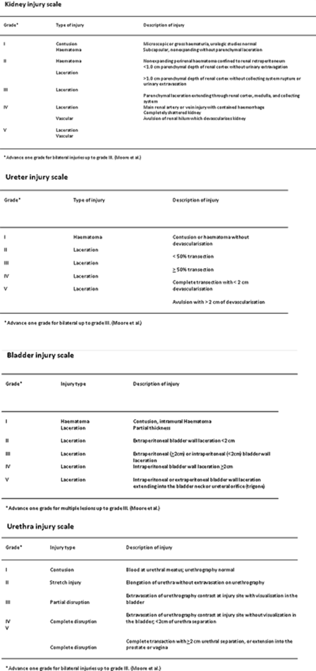
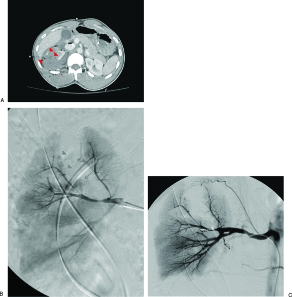
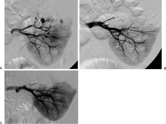
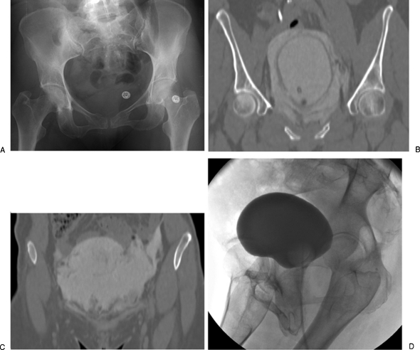
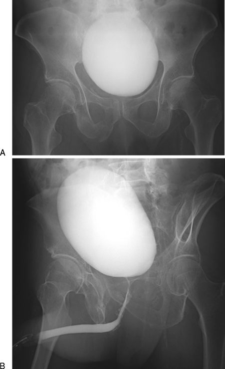
The kidney is the third most common abdominal organ to be injured in trauma, following the spleen and liver, respectively. Several classification systems convey the severity of injury to kidneys, ureter, bladder, and urethra. The most commonly used classification scheme is the American Association for the Surgery of Trauma (AAST) classification of blunt renal injuries, which grades renal injury according the size of laceration and its proximity to the renal hilum. Ureteral injury is graded according to its extent relative to the circumference of the ureter and the extent of associated devascularization. Bladder injury is graded according to its location relative to the peritoneum. Urethral injury is graded according to the extent of damage to surrounding anatomic structures. Although these classification schema may not be always used in common parlance, they do help delineate most important features of urologic tract injury that impact patient management and interventions.

Keywords: Kidney; embolization; genitourinary; interventional radiology; trauma.


Figures

Figure 1
Figure 1
Synopsis of the grading scales for kidney, ureter, urinary bladder, and urethral traumatic injuries.
Figure 2
Figure 2
Embolization of the right posterior segmental artery. (A) Computed tomography (CT) examination of a patient with a penetrating gunshot wound to the right flank shows a grade 4 laceration to the right kidney with multiple foci of active extravasation (arrowheads) as well as a comminuted right posterior 12th rib fracture (arrow). (B) Angiogram demonstrates active extravasation from a superior branch of the right posterior segmental renal artery (arrow), a large wedge-shaped defect corresponding to the renal laceration identified on CT, as well as tiny pseudoaneurysms abutting the margin of the laceration (arrowheads). (C) Successful coil embolization of the right posterior segmental renal artery. Focus of extravasation has largely resolved.
Figure 3
Figure 3
Embolization of a left superior arterial pseudoaneurysm. (A) A 32-year-old woman underwent a partial left nephrectomy one week prior to the diagnostic angiogram that demonstrates at least three pseudoaneurysms arising from the left superior arterial branch supplying the resection margin. (B) Left superior renal artery was successfully embolized. (C) Follow-up angiogram was performed 2 days after the embolization procedure when the patient presented with dropping hemoglobin and hematocrit levels, though no evidence of extravasation was seen.
Figure 4
Figure 4
Percutaneous nephrostomy catheter placement for urinary diversion in a 56-year-old man presenting with abdominal pain and anuria 2 weeks after renal transplantation. (A) Renal scan demonstrated tracer activity in the Jackson-Pruitt drain indicative of a urine leak. (B) Percutaneous nephrostomy catheter was successfully placed for urinary diversion. (C) Antegrade nephrostogram performed 2 days afterward demonstrates a moderate intraperitoneal leak at the ureteral anastomosis of the transplanted kidney. (D) Percutaneous nephrostomy tube was removed ∼3 months later when the leak was shown to be contained on nephrostography (arrow). (E) Follow-up renal scan 20 months after percutaneous nephrostomy catheter removal shows a defect in the region of the contained leak (arrow), but no extravasation of radiotracer to suggest urine leak.
Figure 5
Figure 5
Urinary bladder rupture in a 52-year-old woman after a motor vehicle collision. (A) Pelvis radiograph demonstrates fractures in the left sacral ala, left ischium, bilateral superior pubic rami, and right inferior pubic ramus. (B) A computed tomography cystogram shows a rupture in the right anterolateral aspect of the urinary bladder wall. (C) Presence of contrast in the right paracolic gutter is indicative of an intraperitoneal bladder rupture. Patient was managed conservatively with placement of a Foley catheter. (D) Follow-up cystogram performed ∼1 month afterwards shows interval resolution of the bladder rupture.
Figure 6
Figure 6
Urethral injury in a 63-year-old man who was struck by a motor vehicle. (A) Pelvis radiograph demonstrates fractures in bilateral superior pubic rami and right inferior pubic ramus. Fracture of the right sacral ala is obscured by contrast in the urinary bladder. (B) Retrograde urethrogram demonstrates contrast extravasation at the bulbomembranous junction, indicative of a urethral injury. The patient was treated conservatively by placement of a Foley catheter, which was removed after ∼2 months when the urinary leak was shown to have resolved.

Comment in

References

    1. Titton R L, Gervais D A, Boland G W, Mueller P R. Renal trauma: radiologic evaluation and percutaneous treatment of nonvascular injuries. AJR Am J Roentgenol. 2002;178(6):1507–1511. - PubMed
    1. Bixby S D, Callahan M J, Taylor G A. Imaging blunt pediatric blunt abdominal trauma. Semin Roentgenol. 2008;43(1):72–82. - PubMed
    1. McAleer I M, Kaplan G W, LoSasso B E. Congenital urinary tract anomalies in pediatric renal trauma patients. J Urol. 2002;168(4 Pt 2):1808–1810. discussion 1810. - PubMed
    1. Lorelli D R, Kralovich K A, Seguin C. The impact of pre-existing end-stage renal disease on survival in acutely injured trauma patients. Am Surg. 2001;67(7):693–696. - PubMed
    1. Patel M S, Malinoski D J, Nguyen X M, Hoyt D B. The impact of select chronic diseases on outcomes after trauma: a study from the National Trauma Data Bank. J Am Coll Surg. 2011;212(1):96–104. - PubMed