. 2012 Jun;13(4):267-77.
doi: 10.2174/138920212800793357.

Collective epithelial and mesenchymal cell migration during gastrulation

Affiliations

Collective epithelial and mesenchymal cell migration during gastrulation

Manli Chuai et al. Curr Genomics. 2012 Jun.

Abstract

Gastrulation, the process that puts the three major germlayers, the ectoderm, mesoderm and endoderm in their correct topological position in the developing embryo, is characterised by extensive highly organised collective cell migration of epithelial and mesenchymal cells. We discuss current knowledge and insights in the mechanisms controlling these cell behaviours during gastrulation in the chick embryo. We discuss several ideas that have been proposed to explain the observed large scale vortex movements of epithelial cells in the epiblast during formation of the primitive streak. We review current insights in the control and execution of the epithelial to mesenchymal transition (EMT) underlying the formation of the hypoblast and the ingression of the mesendoderm cells through the streak. We discuss the mechanisms by which the mesendoderm cells move, the nature and dynamics of the signals that guide these movements, as well as the interplay between signalling and movement that result in tissue patterning and morphogenesis. We argue that instructive cell-cell signaling and directed chemotactic movement responses to these signals are instrumental in the execution of all phases of gastrulation.

Keywords: Development; EMT; FGF signalling.; chemotaxis; chick embryo; ingression.


Figures

Fig. (1). Cell flow in epiblast.
A: Section through epiblast of stage EGXII embryo showing the apical localisation of Phospho-ezrin (green) counter staining with Rhodamine Phaloidin to show the actin cytoskeleton). B: Section showing two HNK1 positive cells in the epiblast (upper layer) as well as positive hypoblast cells (lower layer). C: Hypoblast cells in the anterior of the embryo before fusing to form an epithelial sheet (D). E: Automated track analysis of fluorescent chicken embryo (1.25x magnification). Cell paths are shown as lines that go from green to red, with a total journey time of 10 hours. The primitive streak and dual circular 'quiet regions' are clearly visible. F: Diagram of embryo, green Area Opaca, yellow epiblast. Red dots indicating gradient of cells in epiblast that are HNK1 positive, not that there are more positive cells in the posterior of the embryo than in the anterior. Dark red forming secondary epiblast. Grey arrows indicate cell flow patterns as observed in (E). G: Diagram showing relationship between HNK1 expression, cell division and ingression. When cells do not express HNK1 divide they stay in the epiblast, resulting in expansion of the epiblast (black arrows) when they express HNK1 and divide they ingress to form hypoblast cells leading to a contraction. The gradients in anterior expansion and posterior ingression result in cell flows indicated by the curved grey arrows in (F).
Fig. (2). Cell ingression in the primitive streak.
A: Diagram showing changes occurring during ingression in the streak. A signal secreted by the mesoderm cells (light-blue and yellow) results in apical contraction of these cells by myosin (apical blue line). Simultaneously the cells reduce the strength of E-Cadherin medicated interactions (blue double arrows between cells), break down the basal lamina (redline) and down regulate integrin mediated signalling to the matrix ( (green double arrows), polarise by synthesizing actin at their leading edges (orange) and migrate in the internal space of the embryo in response to repulsive ad attractive guidance factors. B: Diagram showing changes in cells shape of random GFP labelled cells from being columnar in the epiblast to being polarised in the direction of migration of mesoderm cells. C: Section of streak region (white arrow) showing expression of N cadherin. Note that cells in the streak start to express N-Cadherin as do all the mesendoderm cells.
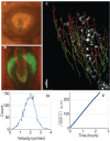
Fig. (3). Migration of GFP labelled mesendoderm cells.
A: Image of a HH3 stage embryo with a small graft of GFP expressing streak cells from a transgenic embryo. B: photograph of the same embryo after 24 hrs of development (HH7) showing that the GFP cells have divided extensively and migrated to form precursors of the blood islands and blood vessels as well as some cells in the posterior of the embryo that have form endoderm. C: Image of tracks of grafted GFP labelled mesoderm cells migrating out of the primitive streak (10 x magnification). Automated track analysis displayed as linear paths coloured from green to red, showing a total journey time of 7 hours. D: Velocity distribution of cells imaged in C. D: Square root of meansquare displacement of cells tracked in (C) showing a linear increase of distance with time showing highly directed migration.
Fig. (4). Signal and cell migration patterns during gastrulation.
Black arrows indicate movement trajectories of mesendoderm cells migrating out of the streak at various anterior to posterior positions in. Cells move out of the streak under the influences of a repulsive FGF8 signal, anterior mesendoderm cells move back in response to FGF4 produced by the forming head process and notochord. Cells in the posterior streak move in the extra-embryonic area to form blood islands response to VEGF. PDGF signalling in the epiblast controls N-Cadherin expression of migrating mesoderm cells and lays out a migration domain for mesoderm cells.

References

    1. Stern CD. In: Gastrulation, from cells to embryo's. Stern CD, editor. New York: Cold Spring Harbor Laboratory Press; 2004.
    1. Chuai M, Dormann D, Weijer CJ. Imaging cell signalling and movement in development. Semin. Cell Dev. Biol. 2009;20(8):947–955. - PubMed
    1. Dormann D, Weijer CJ. Imaging of cell migration. EMBO J. 2006;25(15):3480–93. - PMC - PubMed
    1. Eyal-Giladi H, Kochav S. From cleavage to primitive streak formation: a complementary normal table and a new look at the first stages of the development of the chick. I. General morphology. Dev. Biol. 1976;49(2):321–337. - PubMed
    1. Downie JR. The mechanism of chick blastoderm expansion. J. Embryol. Exp. Morphol. 1976;35(3):559–575. - PubMed

LinkOut - more resources