

Effects of the proapoptotic regulator Bcl-2/adenovirus EIB 19-kDa-interacting protein 3 on the chemosensitivity of human colon cancer cell lines
- PMID: 23205118
- PMCID: PMC3506722
- DOI: 10.3892/ol.2012.933
Effects of the proapoptotic regulator Bcl-2/adenovirus EIB 19-kDa-interacting protein 3 on the chemosensitivity of human colon cancer cell lines
Abstract
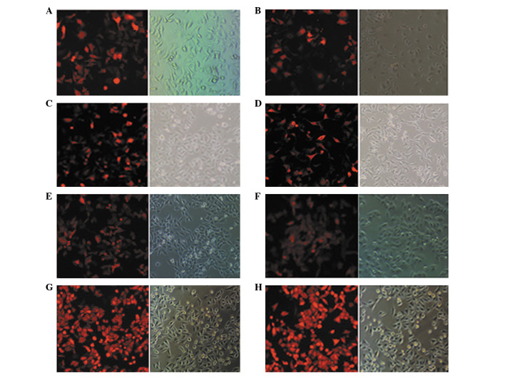
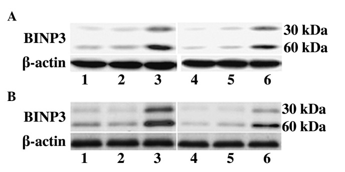
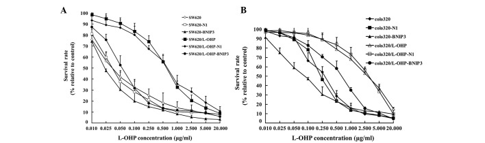
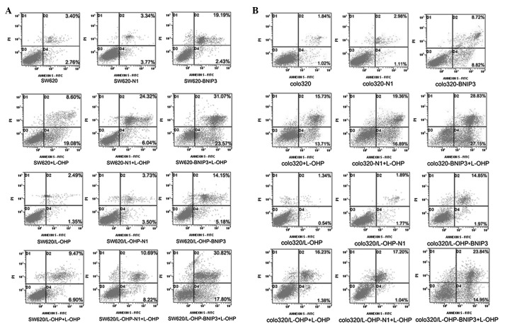
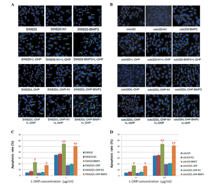
In the clinical setting, drug resistance remains a significant obstacle for successful chemotherapy. Bcl-2/adenovirus EIB 19-kDa-interacting protein 3 (BNIP3) is a proapoptotic member of the Bcl-2 family. To address its potential as a therapeutic target for chemosensitisation, this study investigated the effect of BNIP3 expression on chemosensitivity and reversal of oxaliplatin (L-OHP) resistance in human colon cancer cell lines. A plasmid expressing the BNIP3 gene was transfected into human parental colon cancer cell lines (SW620 and colo320) and L-OHP-resistant colon cancer cell lines (SW620/L-OHP and colo320/L-OHP) using Lipofectamine™ 2000, and the transfection efficiency was determined using fluorescence optics. Western blot analysis identified that SW620/L-OHP and colo320/L-OHP cells expressed lower levels of BNIP3 protein compared with the SW620 and colo320 cells. Transfection with the recombinant BNIP3 plasmid revealed an increase in BNIP3 expression in tumour cells. Following transfection with pDsRed-BNIP3, the chemosensitivity of parental and L-OHP-resistant cell lines to L-OHP was increased (P<0.01), as detected by the Cell Counting Kit-8 (CCK8) assay. Hoechst 33342 staining and flow cytometry revealed that the effects on L-OHP-induced apoptosis were enhanced by the overexpression of BNIP3. Chemosensitisation in human colon cancer cells was observed following treatment with the recombinant BNIP3 plasmid in vitro. The results of this study suggest that BNIP3 is a potential therapeutic target for reversing the resistance of L-OHP-resistant colon cancer cells to L-OHP.
Figures
Similar articles
-
[Effect and mechanism of DNMT inhibitor on the reversal of multidrug resistance in human colon cancer cell line sw620/L-OHP].Sichuan Da Xue Xue Bao Yi Xue Ban. 2010 Nov;41(6):975-9. Sichuan Da Xue Xue Bao Yi Xue Ban. 2010. PMID: 21265097 Chinese.
-
Effects of the proapoptotic regulator Bcl2/adenovirus EIB 19 kDa-interacting protein 3 on radiosensitivity of cervical cancer.Cancer Biother Radiopharm. 2011 Jun;26(3):279-86. doi: 10.1089/cbr.2010.0898. Epub 2011 Jun 28. Cancer Biother Radiopharm. 2011. PMID: 21711117
-
Establishment and biological characteristics of oxaliplatin-resistant human colon cancer cell lines.Chin J Cancer. 2010 Jul;29(7):661-7. doi: 10.5732/cjc.009.10666. Chin J Cancer. 2010. PMID: 20591218
-
The role of autophagy in the treatment of colon cancer by chlorin e6 photodynamic therapy combined with oxaliplatin.Photodiagnosis Photodyn Ther. 2022 Dec;40:103082. doi: 10.1016/j.pdpdt.2022.103082. Epub 2022 Aug 24. Photodiagnosis Photodyn Ther. 2022. PMID: 36028170
-
[The effect of small GTPase Cdc42 on the multidrug resistance of oxaliplatin-resistant colon cancer cell lines].Sichuan Da Xue Xue Bao Yi Xue Ban. 2011 Jul;42(4):466-70. Sichuan Da Xue Xue Bao Yi Xue Ban. 2011. PMID: 21866627 Chinese.
Cited by
-
Implications of Bit1 and AIF overexpressions in esophageal squamous cell carcinoma.Tumour Biol. 2014 Jan;35(1):519-27. doi: 10.1007/s13277-013-1073-8. Epub 2013 Aug 17. Tumour Biol. 2014. PMID: 23955799
References
-
- Jemal A, Bray F, Center MM, Ferlay J, Ward E, Forman D. Global cancer statistics. CA Cancer J Clin. 2011;61:69–90. - PubMed
-
- Segal NH, Saltz LB. Evolving treatment of advanced colon cancer. Annu Rev Med. 2009;60:207–219. - PubMed
-
- Tournigand C, André T, Achille E, Lledo G, Flesh M, Mery-Mignard D, Quinaux E, Couteau C, Buyse M, Ganem G, Landi B, Colin P, Louvet C, de Gramont A. FOLFIRI followed by FOLFOX6 or the reverse sequence in advanced colorectal cancer: a randomized GERCOR study. J Clin Oncol. 2004;22:229–237. - PubMed
-
- Wu DL, Huang F, Lu HZ. Drug-resistant proteins in breast cancer: recent progress in multidrug resistance. Chin J Cancer. 2003;22:441–444. - PubMed
LinkOut - more resources
Full Text Sources