

Developmental dysplasia of the hip: impact of sonographic newborn hip screening on the outcome of early treated decentered hip joints-a single center retrospective comparative cohort study based on Graf's method of hip ultrasonography
- PMID: 23205143
- PMCID: PMC3221760
- DOI: 10.1007/s11832-011-0366-y
Developmental dysplasia of the hip: impact of sonographic newborn hip screening on the outcome of early treated decentered hip joints-a single center retrospective comparative cohort study based on Graf's method of hip ultrasonography
Abstract
PURPOSE/BACKGROUND/INTRODUCTION: The aim of this study was to retrospectively evaluate the impact of neonatal sonographic hip screening using Graf's method for the management and outcome of orthopaedic treatment of decentered hip joints with developmental dysplasia of the hip (DDH), using three decades (1978-2007) of clinical information compiled in a medical database.
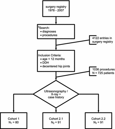
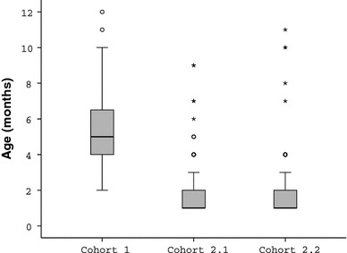
Methods: Three representative cohorts of consecutive cases of decentered hip joints were selected according to different search criteria and inclusion and exclusion parameters: (1) cohort 1 (1978-1982; n = 80), without sonographic screening; (2) cohort 2.1 (1994-1996; n = 91), with nationwide established general sonographic screening according to the Graf-method; (3) cohort 2.2 (2003-2005; n = 91), with sonographic screening including referred cases for open reduction from non-screened populations. These three cohorts were compared for the following parameters: age at initial treatment, successful closed reduction, necessary overhead traction, necessary adductor-tenotomy, rate of open reduction, rate of avascular necrosis (AVN) and rate of secondary acetabuloplasty.
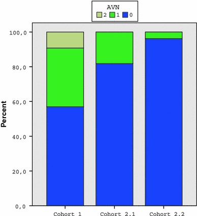
Results: The age at initial treatment was reduced from 5.5 months in the first cohort to 2 months in the two subsequent two cohorts and the rate of successful closed reduction increased from 88.7 to 98.9 and 95.6%, respectively. There was a statistically significant improvement in six out of seven parameters with sonographic hip screening; only the rate of secondary acetabuloplasty did not improve significantly.
Conclusion: Compared to the era before the institution of a sonographic hip screening programme according to the Graf-method in Austria in 1992, ultrasound screening based-treatment of decentered hip joints has become safer, shorter and simpler: "safer" means lower rate of AVN, "shorter" means less treatment time due to earlier onset and "simpler" means that the devices are now less invasive and highly standardized.
Keywords: Decentered hip joints; Developmental dysplasia of the hip; Outcome of treatment; Retrospective comparative cohort study; Sonographic hip screening.
Figures
References
-
- Boeree NR, Clarke NMP. Ultrasound imaging and secondary screening for congenital dislocation of the hip. J Bone Jt Surg (Br) 1994;76-B:525–533. - PubMed
LinkOut - more resources
Full Text Sources
Research Materials
