

Residual hip dysplasia as a risk factor for osteoarthritis in 45 years follow-up of late-detected hip dislocation
- PMID: 23205144
- PMCID: PMC3221757
- DOI: 10.1007/s11832-011-0370-2
Residual hip dysplasia as a risk factor for osteoarthritis in 45 years follow-up of late-detected hip dislocation
Abstract
Purpose: The aim of the study was to assess the role of residual hip dysplasia as a risk factor for osteoarthritis (OA) in developmental dysplasia of the hip (DDH).
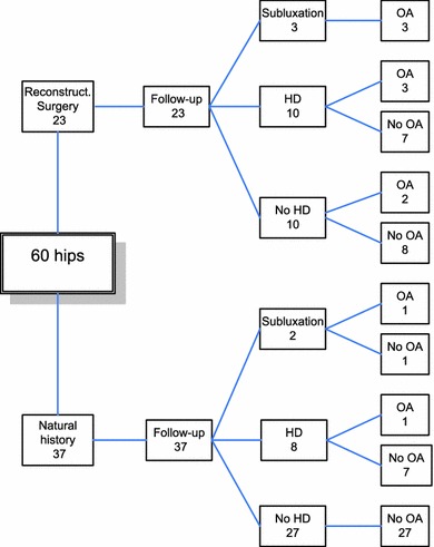
Methods: Fifty-one patients (60 hips) with late-detected DDH were studied. Reduction had been performed at a mean age of 19 months (range 4-65 months). On radiographs at age 8-10 years, at skeletal maturity, and at long-term follow-up, femoral head coverage was assessed using the migration percentage (MP) and centre-edge (CE) angle. OA was diagnosed if the minimum joint space width of the upper part of the joint was <2.0 mm.
Results: The mean age at the last follow-up was 45 years (range 43-49 years) in patients who had not undergone total hip replacement (THR). Ten patients had developed OA and eight of them had undergone THR at a mean age of 40 years (range 32-47 years). There was a clear association between OA and residual hip dysplasia. At the last follow-up, 37 hips had normal CE angles (20° or higher) and OA had developed in only two of them (5%; 95% confidence interval [CI] 1-18%). Hip dysplasia without subluxation (CE angle 10-19°) was seen in 18 hips, of which 14 hips had good outcome and four had OA (22%; 95% CI 6-48%). Subluxation occurred in five hips, of which one had a good long-term outcome and four had OA (80%; 95% CI 28-99%). In patients without late reconstructive surgery, MP increased from the age of 10 years to skeletal maturity; thereafter, no significant change occurred. The CE angle did not change significantly between the age of 10 years and the last follow-up.
Conclusion: Hip dysplasia without subluxation has a relatively good long-term prognosis. Subluxation is a risk factor for osteoarthritis. Thus, children with MP above 33% and CE angle under 10° should be evaluated for reconstructive surgery in order to improve the long-term outcome.
Keywords: Developmental hip dislocation; Long-term follow-up; Osteoarthritis of the hip; Residual hip dysplasia.
Figures
References
-
- Wiberg G. Studies on dysplastic acetabula and congenital subluxation of the hip joint. Acta Chir Scand. 1939;83(Suppl 58):7–135.
-
- Stulberg SD, Harris WH (1974) Acetabular dysplasia and development of osteoarthritis of the hip. In: Harris WH (ed) The hip. Proceedings of the Second Open Scientific Meeting of the Hip Society. Mosby, St. Louis, MO, pp 82–93
LinkOut - more resources
Full Text Sources
Research Materials
