

Relative difference in orbital volume as an indication for surgical reconstruction in isolated orbital floor fractures
- PMID: 23205172
- PMCID: PMC3314253
- DOI: 10.1055/s-0031-1286117
Relative difference in orbital volume as an indication for surgical reconstruction in isolated orbital floor fractures
Abstract
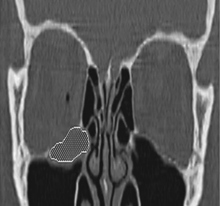
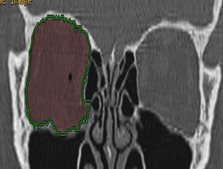
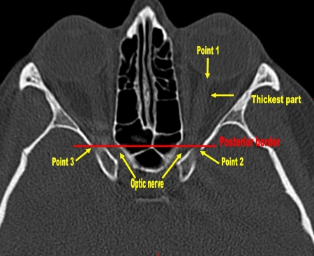
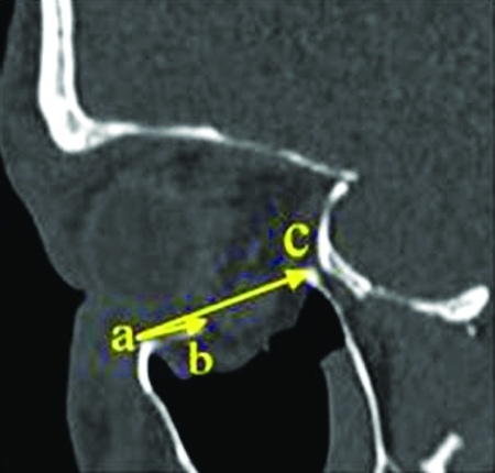
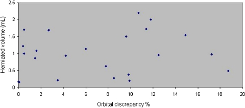
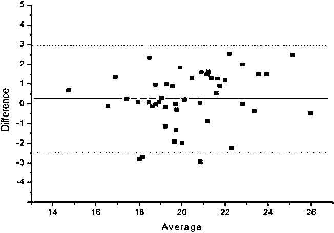
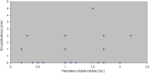
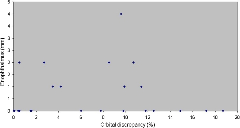
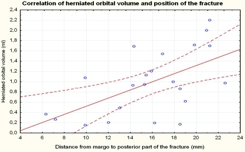
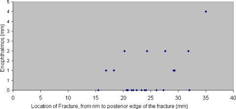
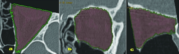
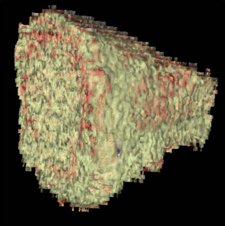
In orbital floor fractures, the estimation of the herniated orbital content in the maxillary sinus has traditionally been the dividing line between surgical and nonsurgical management. In this study, we evaluated whether a relative change in volume would function as an indicator for surgical versus nonsurgical treatment of orbital floor fractures. This was a follow-up study in patients with untreated unilateral isolated orbital floor fractures admitted to our department from March 2003 to April 2007. Patients were contacted by regular mail and invited to have a clinical eye examination. The volume of the orbital content was calculated digitally from the patients' computed tomography scans at the time of their injury. Eighteen subjects with no facial skeleton fracture were included for reference of orbital content volumes. Five of 23 patients showed 2 to 4 mm of enophthalmos, and only three of them had intermittent diplopia. No statistical correlation was found between the herniated volume and enophthalmos. No statistical correlation supporting the supposition that 1 mL of herniated orbital content would result in 1 mm of enophthalmos was found. The relative volume change between the fractured and nonfractured orbit in an individual does not appear to be a useful criterion for surgery. The importance of the herniated orbital tissue for the development of enophthalmos is unclear.
Keywords: Orbital floor fracture; blowout fracture; nonsurgical treatment; orbital volume.
Figures

References
-
- Manson P N, Grivas A, Rosenbaum A, Vannier M, Zinreich J, Iliff N. Studies on enophthalmos: II. The measurement of orbital injuries and their treatment by quantitative computed tomography. Plast Reconstr Surg. 1986;77:203–214. - PubMed
-
- Lee J W, Chiu H Y. Quantitative computed tomography for evaluation of orbital volume change in blow-out fractures. J Formos Med Assoc. 1993;92:349–355. - PubMed
-
- Tong L, Bauer R J, Buchman S R. A current 10-year retrospective survey of 199 surgically treated orbital floor fractures in a nonurban tertiary care center. Plast Reconstr Surg. 2001;108:612–621. - PubMed
-
- Bite U, Jackson I T, Forbes G S, Gehring D G. Orbital volume measurements in enophthalmos using three-dimensional CT imaging. Plast Reconstr Surg. 1985;75:502–508. - PubMed
-
- Koornneef L. Current concepts on the management of orbital blow-out fractures. Ann Plast Surg. 1982;9:185–200. - PubMed
LinkOut - more resources
Full Text Sources
Miscellaneous