. 2013 Mar 15;22(6):975-84.
doi: 10.1089/scd.2012.0438. Epub 2013 Jan 11.

A soluble granulocyte colony stimulating factor decoy receptor as a novel tool to increase hematopoietic cell homing and reconstitution in mice

Affiliations

A soluble granulocyte colony stimulating factor decoy receptor as a novel tool to increase hematopoietic cell homing and reconstitution in mice

Audrey Fortin et al. Stem Cells Dev. .

Abstract

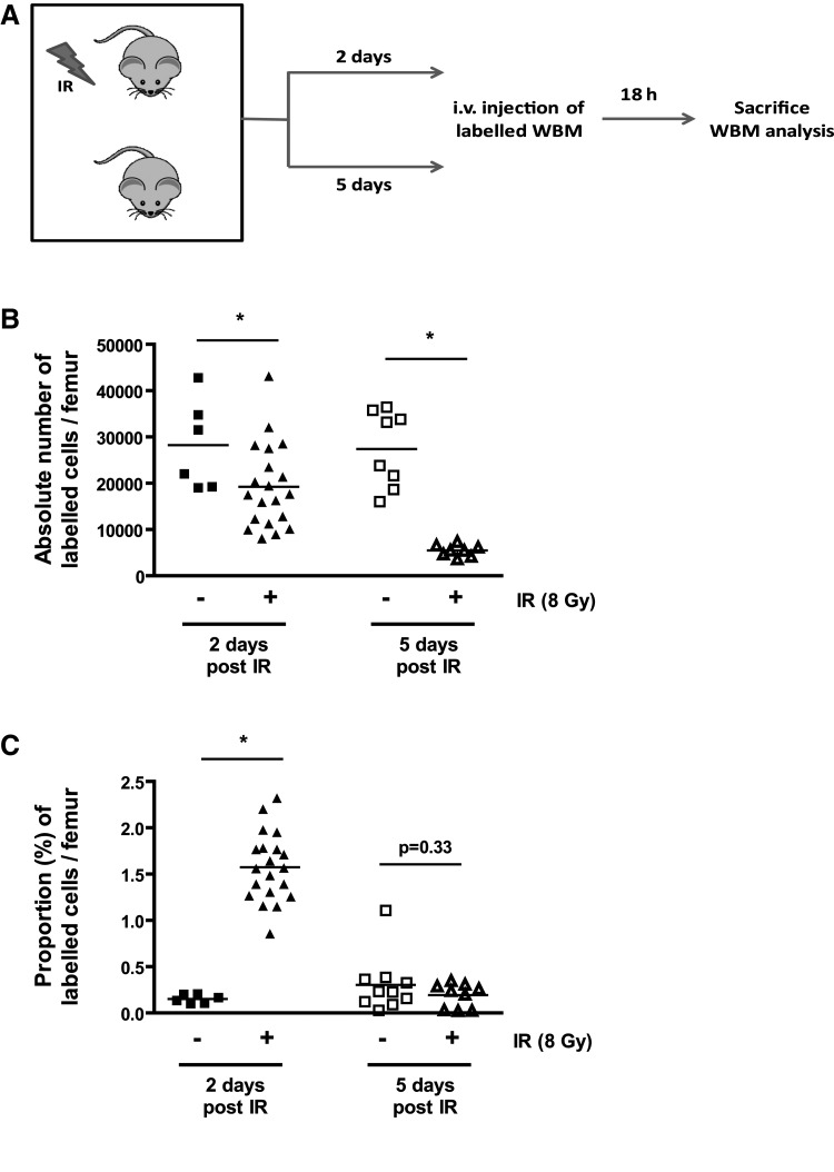
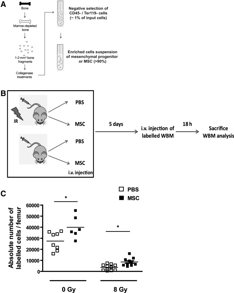
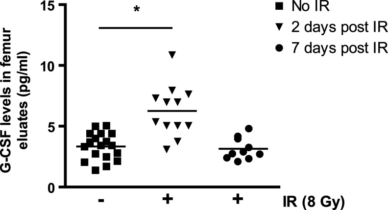
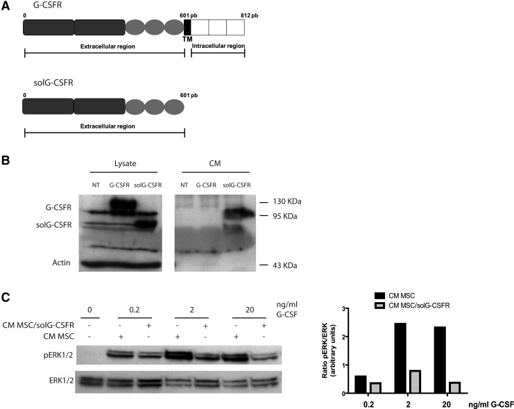
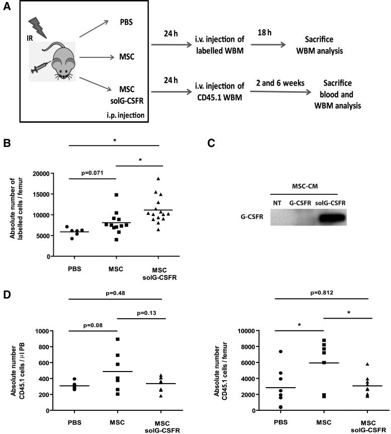
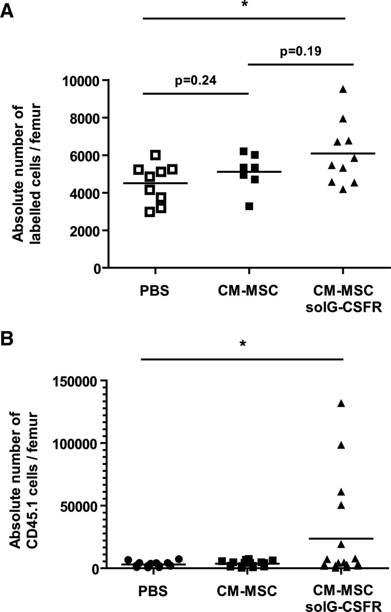
The relative ineffectiveness of hematopoietic stem cells in reaching the bone marrow upon transplantation combined with the limited number of these cells available is a major reason for graft failure and delayed hematopoietic recovery. Hence, the development of strategies that could enhance homing is of high interest. Here, we provide evidence that homing is severely impaired postexposure to ionizing radiation (IR) in mice, an effect we found was time dependent and could be partially rescued using mesenchymal stromal cell (MSC) therapy. In an attempt to further increase homing, we took advantage of our observation that the granulocyte colony stimulating factor (G-CSF), a cytokine known to induce cell mobilization, is increased in the marrow of mice shortly after their exposure to IR. As such, we developed a truncated, yet functional, soluble G-CSF receptor (solG-CSFR), which we hypothesized could act as a decoy and foster homing. Using MSCs or conditioned media as delivery vehicles, we show that an engineered solG-CSFR has the potential to increase homing and hematopoietic reconstitution in mice. Altogether, our results provide novel findings at the interplay of IR and stromal cell therapy and present the regulation of endogenous G-CSF as an innovative proof-of-concept strategy to manipulate hematopoietic cell homing.


Figures

FIG. 1.
FIG. 1.
Impaired homing following exposure to ionizing radiation (IR) is time dependent. (A) Schematic of the experimental procedure. Mice were irradiated or not at the dose of 8 Gy and left untreated for 2 or 5 days after which homing was determined. (B) Absolute number of PKH26+ bone marrow cells present in the femurs of mice at 2 and 5 days post their exposure (+) or not (−) to IR. (C) Same as in B except that the number of PKH26+ bone marrow cells is expressed in proportion to the number of nucleated cell present in the femur at the time of sacrifice. n=6–20, each symbol representing an individual mouse. Student t tests *P<0.05 are shown.
FIG. 2.
FIG. 2.
Injection of mesenchymal stromal cells (MSCs) partially restores homing post-IR. (A) Illustration of the procedure used to isolate MSCs from marrow depleted bones resulting on average in >100-fold enrichment over a nonselected population. (B) Schematic of the experimental design used to determined homing in mice that were exposed (+) or not (−) to IR (8 Gy) and immediately after injected intravenously (i.v.) with 5×10⁴ MSCs or phosphate-buffered saline (PBS) alone. Mice were allowed a 5-day period to allow for the niche regeneration before homing was determined. (C) Homing was determined by counting by flow cytometry the absolute number of PKH26+ bone marrow cells in the femurs of mice. n=8–12, each symbol representing an individual mouse. Student t tests *P<0.05 are shown.
FIG. 3.
FIG. 3.
Increased level of granulocyte colony stimulating factor (G-CSF) in the bone marrow of irradiated mice. G-CSF levels present in the marrow eluates (see methods) collected 2 days and 1 week postexposure of mice to IR (8 Gy) and compared to levels in control (CTR) nonirradiated mice. n=10–18 femur eluates per group. One-way ANOVA test *P<0.05.
FIG. 4.
FIG. 4.
Generation of a soluble G-CSF decoy receptor. (A) Schematic of the G-CSF receptor and its truncated soluble version lacking the transmembrane and extracellular domains. TM=transmembrane domain. (B) Western blot analysis of G-CSF receptor expression in cell lysates and conditioned media (CM) of 293T cells that were either nontransfected (NT), transfected with the full length G-CSFR receptor (G-CSFR) or the soluble G-CSF decoy receptor (solG-CSFR). (C) Western blot analysis of pERK1/2 and total ERK1/2 expression in cell lysates of cultured murine MSCs stimulated with the indicated amount of G-CSF in presence (+) or absence (−) of CM collected from nontransduced MSCs or MSCs expressing the solG-CSFR. Also shown is the inhibition of pERK by CM collected from MSCs expressing the solG-CSFR as determined by the ratio of pERK/ERK in arbitrary units. The bar graph shown is representative of n=3 individual blots.
FIG. 5.
FIG. 5.
Altered homing and engraftment by the secretion of a soluble G-CSF decoy receptor. (A) Schematic of the experiment. In brief, CD45.2 mice were sublethally irradiated at the dose of 8 Gy and immediately injected intraperitoneally (i.p.) with PBS alone or 15×106 MSCs modified or not to secrete the solG-CSFR. Twenty-four hours later, mice were injected intravenously (i.v.) with 2×10⁶ CD45.1 or 5×10⁶ unfractionated whole bone marrow (WBM) cells labeled with CFSE. (B) Shown is the absolute number of fluorescently labeled cells present in bone marrow (femurs) at the time of sacrifice as determined by flow cytometry. n=6–15 mice per group, each symbol representing an individual mouse. (C) The presence of the solG-CSFR in the CM of stably transduced cultured MSCs was confirmed by western blot (NT=not transduced). (D) Engraftment levels in blood and bone marrow were determined at 13 and 45 days, respectively, following the injection of WBM cells. Absolute counts of CD45.1 cells were determined by flow cytometry. n=7–9 mice per group, each symbol representing an individual mouse. Student t tests *P<0.05 are shown.
FIG. 6.
FIG. 6.
Injection of CM collected from MSCs is sufficient to alter homing and hematopoietic reconstitution. We used the same experimental design as in figure 5 except that conditioned media collected from MSCs (CM-MSCs) or MSCs secreting the solG-CSFR (CM-MSCs solG-CSRR) were injected before the injection of 2×10⁵ CD45.1 or 5×10⁶ unfractionated whole bone marrow (WBM) cells labeled with CFSE. (A) Shown is the absolute number of fluorescently labeled cells present in bone marrow (femurs) at the time of sacrifice as determined by flow cytometry. n=7–10 mice per group, each symbol representing an individual mouse. (B) Hematopoietic reconstitution levels in bone marrow were determined 1 week following the injection of CD45.1 cells. n=14–17 mice per group, each symbol representing an individual mouse. Student t tests *P<0.05 are shown.

References

    1. Rocha V. Broxmeyer HE. New approaches for improving engraftment after cord blood transplantation. Biol Blood Marrow Transplant. 2010;16:S126–S132. - PubMed
    1. Jetmore A. Plett PA. Tong X. Wolber FM. Breese R. Abonour R. Orschell-Traycoff CM. Srour EF. Homing efficiency, cell cycle kinetics, and survival of quiescent and cycling human CD34(+) cells transplanted into conditioned NOD/SCID recipients. Blood. 2002;99:1585–1593. - PubMed
    1. Petit I. Szyper-Kravitz M. Nagler A. Lahav M. Peled A. Habler L. Ponomaryov T. Taichman RS. Arenzana-Seisdedos F, et al. G-CSF induces stem cell mobilization by decreasing bone marrow SDF-1 and up-regulating CXCR4. Nat Immunol. 2002;3:687–694. - PubMed
    1. Sato N. Sawada K. Takahashi TA. Mogi Y. Asano S. Koike T. Sekiguchi S. A time course study for optimal harvest of peripheral blood progenitor cells by granulocyte colony-stimulating factor in healthy volunteers. Exp Hematol. 1994;22:973–978. - PubMed
    1. Aiuti A. Webb IJ. Bleul C. Springer T. Gutierrez-Ramos JC. The chemokine SDF-1 is a chemoattractant for human CD34+ hematopoietic progenitor cells and provides a new mechanism to explain the mobilization of CD34+ progenitors to peripheral blood. J Exp Med. 1997;185:111–120. - PMC - PubMed

Publication types

MeSH terms

Substances

LinkOut - more resources