

DUX4 expression in FSHD muscle cells: how could such a rare protein cause a myopathy?
- PMID: 23206257
- PMCID: PMC3823138
- DOI: 10.1111/j.1582-4934.2012.01647.x
DUX4 expression in FSHD muscle cells: how could such a rare protein cause a myopathy?
Abstract
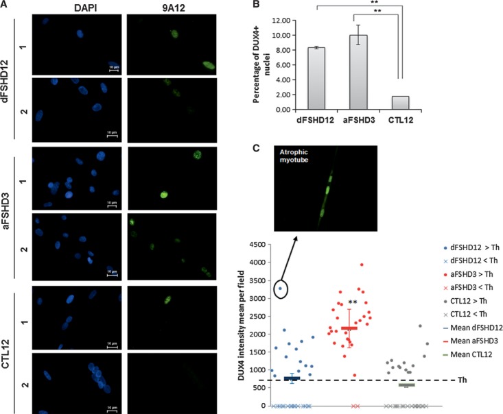
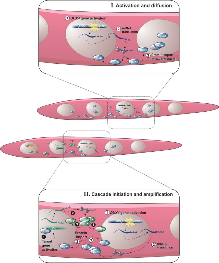
Facioscapulohumeral muscular dystrophy (FSHD) is one of the most frequent hereditary muscle disorders. It is linked to contractions of the D4Z4 repeat array in 4q35. We have characterized the double homeobox 4 (DUX4) gene in D4Z4 and its mRNA transcribed from the distal D4Z4 unit to a polyadenylation signal in the flanking pLAM region. It encodes a transcription factor expressed in FSHD but not healthy muscle cells which initiates a gene deregulation cascade causing differentiation defects, muscle atrophy and oxidative stress. PITX1 was the first identified DUX4 target and encodes a transcription factor involved in muscle atrophy. DUX4 was found expressed in only 1/1000 FSHD myoblasts. We have now shown it was induced upon differentiation and detected in about 1/200 myotube nuclei. The DUX4 and PITX1 proteins presented staining gradients in consecutive myonuclei which suggested a diffusion as known for other muscle nuclear proteins. Both protein half-lifes were regulated by the ubiquitin-proteasome pathway. In addition, we could immunodetect the DUX4 protein in FSHD muscle extracts. As a model, we propose the DUX4 gene is stochastically activated in a small number of FSHD myonuclei. The resulting mRNAs are translated in the cytoplasm around an activated nucleus and the DUX4 proteins diffuse to adjacent nuclei where they activate target genes such as PITX1. The PITX1 protein can further diffuse to additional myonuclei and expand the transcriptional deregulation cascade initiated by DUX4. Together the diffusion and the deregulation cascade would explain how a rare protein could cause the muscle defects observed in FSHD.
© 2012 The Authors. Published by Foundation for Cellular and Molecular Medicine/Blackwell Publishing Ltd. This is an open access article under the terms of the Creative Commons Attribution License, which permits use, distribution and reproduction in any medium, provided the original work is properly cited.
Figures


Similar articles
-
The FSHD atrophic myotube phenotype is caused by DUX4 expression.PLoS One. 2011;6(10):e26820. doi: 10.1371/journal.pone.0026820. Epub 2011 Oct 28. PLoS One. 2011. PMID: 22053214 Free PMC article.
-
Overexpression of the double homeodomain protein DUX4c interferes with myofibrillogenesis and induces clustering of myonuclei.Skelet Muscle. 2018 Jan 12;8(1):2. doi: 10.1186/s13395-017-0148-4. Skelet Muscle. 2018. PMID: 29329560 Free PMC article.
-
DUX4, a candidate gene of facioscapulohumeral muscular dystrophy, encodes a transcriptional activator of PITX1.Proc Natl Acad Sci U S A. 2007 Nov 13;104(46):18157-62. doi: 10.1073/pnas.0708659104. Epub 2007 Nov 5. Proc Natl Acad Sci U S A. 2007. PMID: 17984056 Free PMC article.
-
A complex interplay of genetic and epigenetic events leads to abnormal expression of the DUX4 gene in facioscapulohumeral muscular dystrophy.Neuromuscul Disord. 2016 Dec;26(12):844-852. doi: 10.1016/j.nmd.2016.09.015. Epub 2016 Sep 19. Neuromuscul Disord. 2016. PMID: 27816329 Review.
-
Facioscapulohumeral muscular dystrophy (FSHD): an enigma unravelled?Hum Genet. 2012 Mar;131(3):325-40. doi: 10.1007/s00439-011-1100-z. Epub 2011 Oct 9. Hum Genet. 2012. PMID: 21984394 Review.
Cited by
-
Facioscapulohumeral muscular dystrophy: genetics, gene activation and downstream signalling with regard to recent therapeutic approaches: an update.Orphanet J Rare Dis. 2021 Mar 12;16(1):129. doi: 10.1186/s13023-021-01760-1. Orphanet J Rare Dis. 2021. PMID: 33712050 Free PMC article. Review.
-
Genetic Approaches for the Treatment of Facioscapulohumeral Muscular Dystrophy.Front Pharmacol. 2021 Mar 12;12:642858. doi: 10.3389/fphar.2021.642858. eCollection 2021. Front Pharmacol. 2021. PMID: 33776777 Free PMC article. Review.
-
Gene Editing Targeting the DUX4 Polyadenylation Signal: A Therapy for FSHD?J Pers Med. 2020 Dec 23;11(1):7. doi: 10.3390/jpm11010007. J Pers Med. 2020. PMID: 33374516 Free PMC article.
-
All-in-one vectors for epigenetic CRISPR inhibition of DUX4-fl in facioscapulohumeral muscular dystrophy.Mol Ther Methods Clin Dev. 2025 Aug 7;33(3):101546. doi: 10.1016/j.omtm.2025.101546. eCollection 2025 Sep 11. Mol Ther Methods Clin Dev. 2025. PMID: 40893163 Free PMC article.
-
Temporal variation in p38-mediated regulation of DUX4 in facioscapulohumeral muscular dystrophy.Sci Rep. 2024 Nov 2;14(1):26437. doi: 10.1038/s41598-024-77911-8. Sci Rep. 2024. PMID: 39488616 Free PMC article.
References
-
- Hewitt JE, Lyle R, Clark LN, et al. Analysis of the tandem repeat locus D4Z4 associated with facioscapulohumeral muscular dystrophy. Hum Mol Genet. 1994;3:1287–95. - PubMed
-
- Lyle R, Wright TJ, Clark LN, et al. The FSHD-associated repeat, D4Z4, is a member of a dispersed family of homeobox-containing repeats, subsets of which are clustered on the short arms of the acrocentric chromosomes. Genomics. 1995;28:389–97. - PubMed
-
- Wijmenga C, Hewitt JE, Sandkuijl LA, et al. Chromosome 4q DNA rearrangements associated with facioscapulohumeral muscular dystrophy. Nat Genet. 1992;2:26–30. - PubMed
Publication types
MeSH terms
Substances
Grants and funding
LinkOut - more resources
Full Text Sources
Other Literature Sources
Research Materials
