

Placenta-specific novel splice variants of Rho GDP dissociation inhibitor β are highly expressed in cancerous cells
- PMID: 23206989
- PMCID: PMC3554444
- DOI: 10.1186/1756-0500-5-666
Placenta-specific novel splice variants of Rho GDP dissociation inhibitor β are highly expressed in cancerous cells
Abstract
Background: Alternative splicing of pre-mRNA transcripts not only plays a role in normal molecular processes but is also associated with cancer development. While normal transcripts are ubiquitously expressed in normal tissues, splice variants created through abnormal alternative splicing events are often expressed in cancer cells. Although the Rho GDP dissociation inhibitor β (ARHGDIB) gene has been found to be ubiquitously expressed in normal tissues and involved in cancer development, the presence of splice variants of ARHGDIB has not yet been investigated.
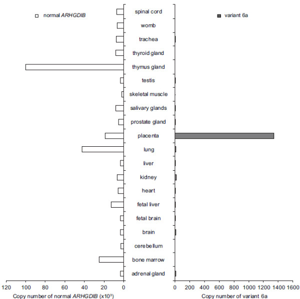
Results: Validation analysis for the presence of and exon structures of splice variants of ARHGDIB, performed using reverse-transcriptase polymerase chain reaction and DNA sequencing, successfully identified novel splice variants of ARHGDIB, that is, 6a, 6b, and 6c, in colon, pancreas, stomach, and breast cancer cell lines. Quantitative real-time polymerase chain reaction analysis showed that these variants were also highly expressed in normal placental tissue but not in other types of normal tissue.
Conclusions: Expression of ARHGDIB variants 6a, 6b, and 6c appears to be restricted to cancer cells and normal placental tissue, suggesting that these variants possess cancer-specific functions and, as such, are potential cancer-related biomarkers.
Figures


Similar articles
-
Systematic investigation of biomarker-like role of ARHGDIB in breast cancer.Cancer Biomark. 2020;28(1):101-110. doi: 10.3233/CBM-190562. Cancer Biomark. 2020. PMID: 32176626
-
Functional classification of DNA variants by hybrid minigenes: Identification of 30 spliceogenic variants of BRCA2 exons 17 and 18.PLoS Genet. 2017 Mar 24;13(3):e1006691. doi: 10.1371/journal.pgen.1006691. eCollection 2017 Mar. PLoS Genet. 2017. PMID: 28339459 Free PMC article.
-
Re-splicing of mature mRNA in cancer cells promotes activation of distant weak alternative splice sites.Nucleic Acids Res. 2012 Sep;40(16):7896-906. doi: 10.1093/nar/gks520. Epub 2012 Jun 6. Nucleic Acids Res. 2012. PMID: 22675076 Free PMC article.
-
Tissue-specific splicing of two mutually exclusive exons of the chicken beta-tropomyosin pre-mRNA: positive and negative regulations.Biochimie. 1996;78(6):457-65. doi: 10.1016/0300-9084(96)84752-3. Biochimie. 1996. PMID: 8915535 Review.
-
Alternative splicing of the estrogen receptor primary transcript normally occurs in estrogen receptor positive tissues and cell lines.J Steroid Biochem Mol Biol. 1996 Jan;56(1-6 Spec No):99-105. doi: 10.1016/0960-0760(95)00227-8. J Steroid Biochem Mol Biol. 1996. PMID: 8603053 Review.
Cited by
-
Cancer survival analysis using semi-supervised learning method based on Cox and AFT models with L1/2 regularization.BMC Med Genomics. 2016 Mar 1;9:11. doi: 10.1186/s12920-016-0169-6. BMC Med Genomics. 2016. PMID: 26932592 Free PMC article.
-
Copy number variation in hereditary non-polyposis colorectal cancer.Genes (Basel). 2013 Sep 26;4(4):536-55. doi: 10.3390/genes4040536. Genes (Basel). 2013. PMID: 24705261 Free PMC article.
References
-
- Becker KF, Atkinson MJ, Reich U, Becker I, Nekarda H, Siewert JR, Hofler H. E-cadherin gene mutations provide clues to diffuse type gastric carcinomas. Cancer Res. 1994;54:3845–3852. - PubMed
-
- Gayther SA, Barski P, Batley SJ, Li L, de Foy KA, Cohen SN, Ponder BA, Caldas C. Aberrant splicing of the TSG101 and FHIT genes occurs frequently in multiple malignancies and in normal tissues and mimics alterations previously described in tumours. Oncogene. 1997;15:2119–2126. doi: 10.1038/sj.onc.1201591. - DOI - PubMed
-
- Barbour AP, Reeder JA, Walsh MD, Fawcett J, Antalis TM, Gotley DC. Expression of the CD44v2-10 isoform confers a metastatic phenotype: importance of the heparan sulfate attachment site CD44v3. Cancer Res. 2003;63:887–892. - PubMed
Publication types
MeSH terms
Substances
LinkOut - more resources
Full Text Sources
Miscellaneous
