. 2013 Feb;33(4):701-11.
doi: 10.1128/MCB.01057-12. Epub 2012 Dec 3.

Cernunnos deficiency reduces thymocyte life span and alters the T cell repertoire in mice and humans

Affiliations

Cernunnos deficiency reduces thymocyte life span and alters the T cell repertoire in mice and humans

Gabriella Vera et al. Mol Cell Biol. 2013 Feb.

Abstract

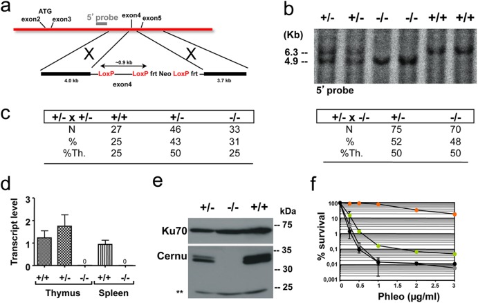
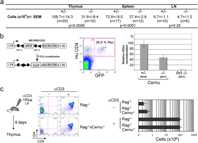
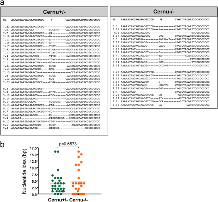
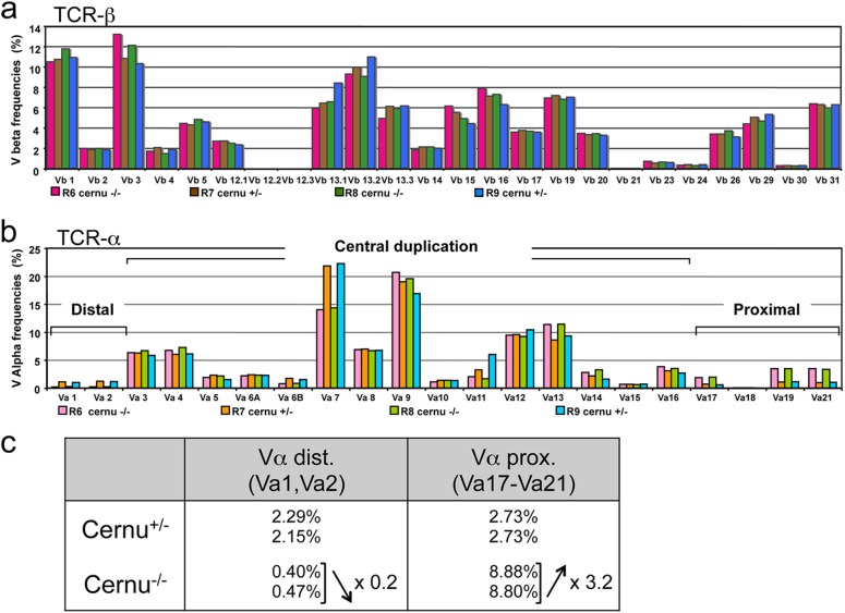
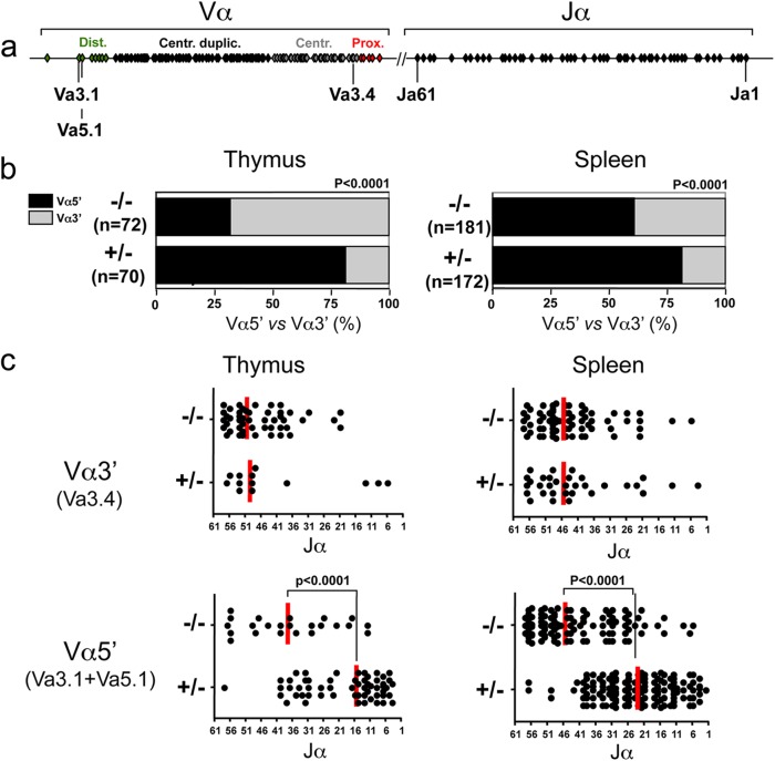
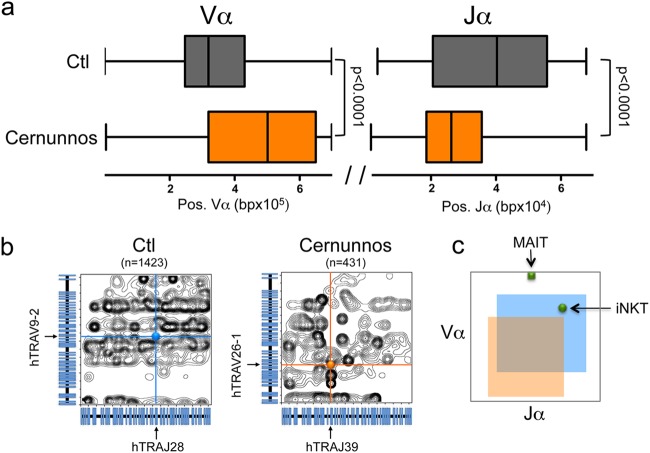
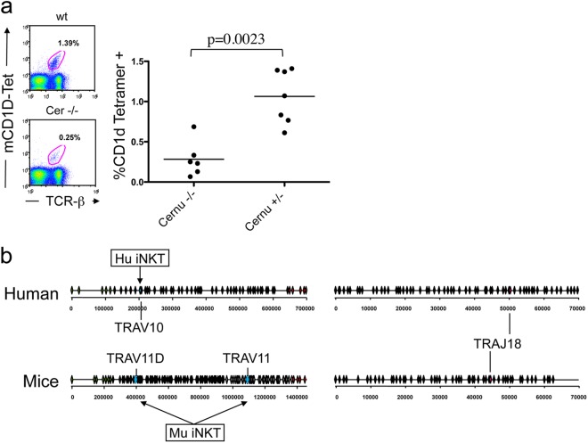
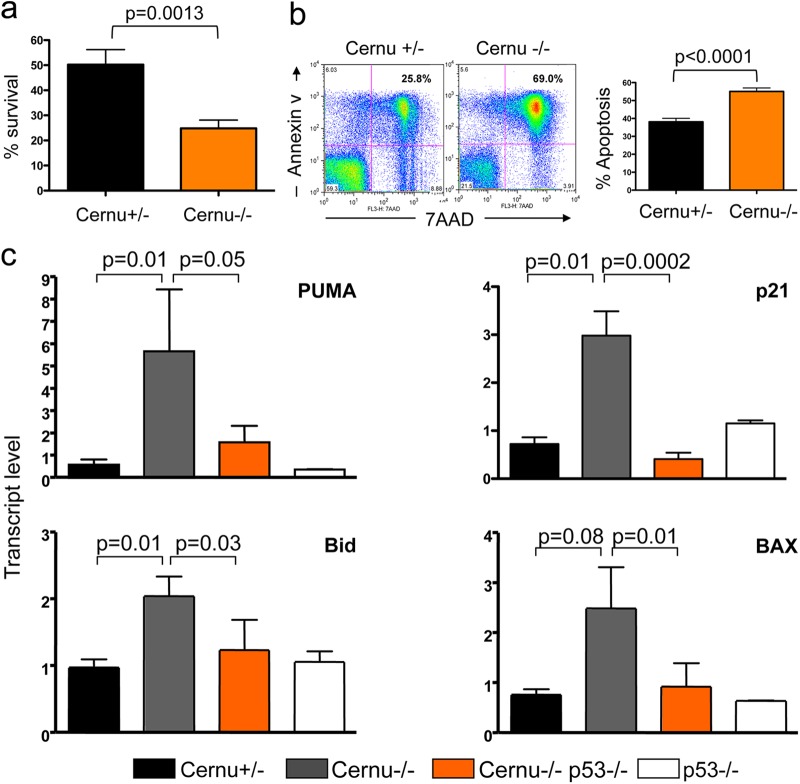
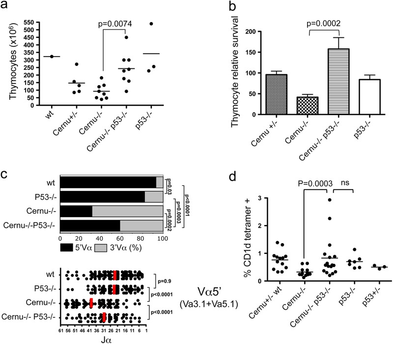
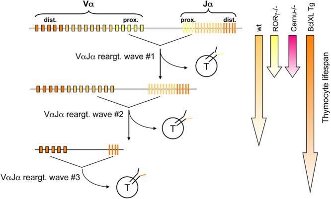
Cernunnos is a DNA repair factor of the nonhomologous end-joining machinery. Its deficiency in humans causes radiosensitive severe combined immune deficiency (SCID) with microcephaly, characterized in part by a profound lymphopenia. In contrast to the human condition, the immune system of Cernunnos knockout (KO) mice is not overwhelmingly affected. In particular, Cernunnos is dispensable during V(D)J recombination in lymphoid cells. Nevertheless, the viability of thymocytes is reduced in Cernunnos KO mice, owing to the chronic activation of a P53-dependent DNA damage response. This translates into a qualitative alteration of the T cell repertoire to one in which the most distal Vα and Jα segments are missing. This results in the contraction of discrete T cell populations, such as invariant natural killer T (iNKT) and mucosa-associated invariant T (MAIT) cells, in both humans and mice.


Figures

Fig 1
Fig 1
Generation of Cernu−/− mice. (a) Schematic representation of the KO strategy and location of the external 5′ probe. Exon 4 was flanked by LoxP sites. (b) Southern blot analysis of tail DNA by use of a 5′ probe. The blot shows the 6.3-kb wt band and the 4.9-kb KO allele on a BclI digest. (c) Mendelian distributions of genotypes in offspring of Cernu+/− × Cernu+/− and Cernu+/− × Cernu−/− intercrosses. %Th., percent theoretical. (d) Quantitative RT-PCR evaluation of Cernunnos transcript levels in thymuses and spleens of +/+, +/−, and −/− littermates. (e) Western blot analysis showing the absence of the 35-kDa Cernunnos protein in MEFs. Anti-Ku-70 antibody was used as a loading control. **, nonspecific band. (f) Phleomycin sensitivity of Cernu−/− (green circles), Xrcc4−/− (gray circles), DNA Lig4−/− (black circles), and wt (orange circles) MEFs. The data represent one of two separate experiments, with values representing the means of two independent determinations for each point.
Fig 2
Fig 2
(a) Lymphocyte cellularity in thymuses, spleens, and lymph nodes (LN) of Cernu+/− and Cernu−/− mice. SEM, standard error of the mean. (b) V(D)J recombination activity in purified CD4 CD8 thymocytes. Transduced cells were identified through expression of the huCD4 gene from the reporter. The % recombination represents the fraction of GFP-positive cells among the huCD4-positive cells. The relative V(D)J activity was calculated by comparison to the level attained in thymocytes from Cernu+/− mice, used as a 100% activity control. (c) Proliferative capacity of thymocytes. Cernu−/− Rag1−/− and Rag1−/− mice were injected i.p. with anti-CD3.
Fig 3
Fig 3
(a) Nucleotide sequence analysis of coding joints recovered from pMX-RSS-12/23-transduced thymocytes from 2 Cernu+/− and 2 Cernu−/− mice. (b) Similar frequencies of nucleotide loss at coding joints recovered from pMX-RSS-12/23-transduced thymocytes from Cernu+/− and Cernu−/− mice.
Fig 4
Fig 4
Immunoscope analysis of TCR-β (a) and TCR-α (b) repertoires from 2 Cernu−/− (R6 and R8) and 2 Cernu+/− (R7 and R9) mice. (c) Differential representation of distal (5′) and proximal (3′) Vα segments in Cernu−/− and Cernu+/− T cells.
Fig 5
Fig 5
(a) Schematic representation of murine TCR Vα and Jα clusters and positions of the three Vα genes coamplified by PCR using a unique set of primers. (b) Differential representation of distal Vαs (mTRAV3.1 and mTRAV5.1) versus proximal Vαs (mTRAV3.4) after cloning and sequencing of RT-PCR products from the thymus and the spleen. (c) TCR Jα representation in distal and proximal Vα-containing transcripts from thymuses and spleens of Cernu−/− and Cernu+/− mice. Vertical red lines correspond to the medians for Jα usage.
Fig 6
Fig 6
TCR-α repertoire in a human Cernunnos patient. (a) Box-and-whisker representation of Vα and Jα usage in TCR-α transcripts from one Cernunnos patient and one control individual as determined by 5′RACE PCR and NGS. Each VαJα transcript (orange and gray areas for the Cernunnos patient and the control individual, respectively) is positioned according to the relative locations (in bp) of its Vα and Jα elements in the genome. (b) Contour plot representation of TCR-α transcripts from a Cernunnos patient (431 sequences) and a control individual (1,423 sequences). Blue and orange bullets represent the VαJα barycenter in each case. (c) Boxes representing 75% of TCR-α transcript sequences for a Cernunnos patient (orange square) and a control individual (blue square). The two green bullets represent the theoretical positions of TCR-α chains from iNKT (hTRAV10, at bp 2.0 × 10⁵; and hTRAJ18, at bp 5.0 × 10⁴) and MAIT (hTRAV1.2, at bp 0.2 × 10⁵; and hTRAJ33, at bp 3.3 × 10⁴) cells, respectively.
Fig 7
Fig 7
(a) Detection of iNKT cells in the spleen by dual staining with anti-TCR-β and CD1d tetramer. (b) Schematic representation of human and murine TCR Vα and Jα clusters, including the locations of TRAV and TRAJ used by iNKT cells. Two Jα segments can be used by murine NKT cells, i.e., TRAV11D and the most downstream segment, TRAV11, because of the duplication of the central TCR Jα segments in mice.
Fig 8
Fig 8
Thymocyte survival (a) and apoptosis (b) in 24-h cultures in vitro. (c) Quantitative RT-PCR analysis of P53 target genes in Cernu−/− thymocytes.
Fig 9
Fig 9
Partial normalization of Cernu−/− thymocyte viability upon P53 inactivation. (a) Total thymocyte counts. (b) Relative thymocyte survival in 24-h cultures. (c) Differential TCR Vα usage in splenic T cells, analyzed using PCR coamplification of two 5′ Vα (Va3.1 and Va5.1) and one 3′ Vα (Va3.4) segment. The vertical red lines represent the medians for Jα usage. (d) iNKT cell recovery in the spleen. ns, not significant.
Fig 10
Fig 10
Multiple waves of TCR-α rearrangements during thymocyte development. DNA accessibility in the TCR Jα cluster is regulated such that the first VαJα rearrangements are targeted to the 5′ side of the TCR Jα cluster. If the thymocytes expressing the resulting TCR are not positively selected, Rag1 and Rag2 expression continues, and a second wave of rearrangement involving upstream Vα and downstream Jα segments occurs. In the absence of positive selection of this newly expressed TCR, a third wave of VαJα recombination occurs, and so on. The possibility of thymocytes undergoing several waves of TCR-α rearrangement depends on their life span. In RORγT−/− mice, thymocyte viability is decreased, resulting in a bias of Jα usage toward the most 5′ elements (first wave). In contrast, the thymocyte life span is increased in BclXL Tg mice, allowing several waves of recombination and the resulting skewing of Jα usage toward the most downstream elements.

References

    1. Helmink BA, Sleckman BP. 2012. The response to and repair of RAG-mediated DNA double-strand breaks. Annu. Rev. Immunol. 30:175–202 - PMC - PubMed
    1. Lieber MR. 2010. The mechanism of double-strand DNA break repair by the nonhomologous DNA end-joining pathway. Annu. Rev. Biochem. 79:181–211 - PMC - PubMed
    1. Buck D, Malivert L, de Chasseval R, Barraud A, Fondaneche MC, Sanal O, Plebani A, Stephan JL, Hufnagel M, le Deist F, Fischer A, Durandy A, de Villartay JP, Revy P. 2006. Cernunnos, a novel nonhomologous end-joining factor, is mutated in human immunodeficiency with microcephaly. Cell 124:287–299 - PubMed
    1. Ahnesorg P, Smith P, Jackson SP. 2006. XLF interacts with the XRCC4-DNA ligase IV complex to promote DNA nonhomologous end-joining. Cell 124:301–313 - PubMed
    1. Andres SN, Modesti M, Tsai CJ, Chu G, Junop MS. 2007. Crystal structure of human XLF: a twist in nonhomologous DNA end-joining. Mol. Cell 28:1093–1101 - PubMed

Publication types

MeSH terms