

MCARD-mediated gene transfer of GRK2 inhibitor in ovine model of acute myocardial infarction
- PMID: 23208013
- PMCID: PMC3695486
- DOI: 10.1007/s12265-012-9418-z
MCARD-mediated gene transfer of GRK2 inhibitor in ovine model of acute myocardial infarction
Abstract
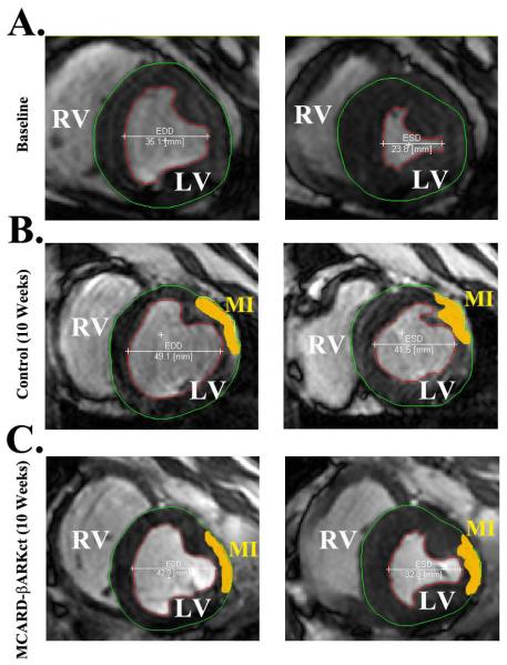
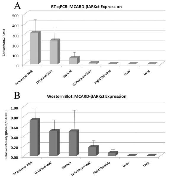
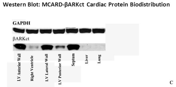
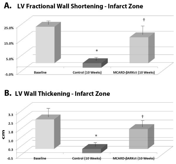
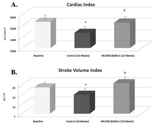
β-Adrenergic receptor (βAR) dysfunction in acute myocardial infarction (MI) is associated with elevated levels of the G-protein-coupled receptor kinase-2 (GRK2), which plays a key role in heart failure progression. Inhibition of GRK2 via expression of a peptide βARKct transferred by molecular cardiac surgery with recirculating delivery (MCARD) may be a promising intervention. Five sheep underwent scAAV6-mediated MCARD delivery of βARKct, and five received no treatment (control). After a 3-week period, the branch of the circumflex artery (OM1) was ligated. Quantitative PCR data showed intense βARKct expression in the left ventricle (LV). Circumferential fractional shortening was 23.4 ± 7.1 % (baseline) vs. -2.9 ± 5.2 % (p < 0.05) in the control at 10 weeks. In the MCARD-βARKct group, this parameter was close to baseline. The same trend was observed with LV wall thickening. Cardiac index fully recovered in the MCARD-βARKct group. LV end-diastolic volume and LV end-diastolic pressure did not differ in both groups. MCARD-mediated βARKct gene expression results in preservation of regional and global systolic function after acute MI without arresting progressive ventricular remodeling.
Figures


Similar articles
-
AAV6-βARKct gene delivery mediated by molecular cardiac surgery with recirculating delivery (MCARD) in sheep results in robust gene expression and increased adrenergic reserve.J Thorac Cardiovasc Surg. 2012 Mar;143(3):720-726.e3. doi: 10.1016/j.jtcvs.2011.08.048. Epub 2011 Dec 3. J Thorac Cardiovasc Surg. 2012. PMID: 22143102 Free PMC article.
-
Safety and efficacy of high-dose adeno-associated virus 9 encoding sarcoplasmic reticulum Ca(2+) adenosine triphosphatase delivered by molecular cardiac surgery with recirculating delivery in ovine ischemic cardiomyopathy.J Thorac Cardiovasc Surg. 2014 Sep;148(3):1065-72, 1073e1-2; discussion1072-3. doi: 10.1016/j.jtcvs.2014.05.070. Epub 2014 Jun 7. J Thorac Cardiovasc Surg. 2014. PMID: 25037619 Free PMC article.
-
AAV6.βARKct cardiac gene therapy ameliorates cardiac function and normalizes the catecholaminergic axis in a clinically relevant large animal heart failure model.Eur Heart J. 2013 May;34(19):1437-47. doi: 10.1093/eurheartj/ehr447. Epub 2012 Jan 19. Eur Heart J. 2013. PMID: 22261894 Free PMC article.
-
GRK2 as a novel gene therapy target in heart failure.J Mol Cell Cardiol. 2011 May;50(5):785-92. doi: 10.1016/j.yjmcc.2010.08.014. Epub 2010 Aug 25. J Mol Cell Cardiol. 2011. PMID: 20800067 Free PMC article. Review.
-
betaARKct: a therapeutic approach for improved adrenergic signaling and function in heart disease.J Cardiovasc Transl Res. 2010 Oct;3(5):499-506. doi: 10.1007/s12265-010-9206-6. Epub 2010 Jul 10. J Cardiovasc Transl Res. 2010. PMID: 20623214 Review.
Cited by
-
AAV-mediated gene therapy for heart failure: enhancing contractility and calcium handling.F1000Prime Rep. 2013 Aug 1;5:27. doi: 10.12703/P5-27. eCollection 2013. F1000Prime Rep. 2013. PMID: 23967378 Free PMC article.
-
Targeting GPCR-Gβγ-GRK2 signaling as a novel strategy for treating cardiorenal pathologies.Biochim Biophys Acta Mol Basis Dis. 2017 Aug;1863(8):1883-1892. doi: 10.1016/j.bbadis.2017.01.020. Epub 2017 Jan 25. Biochim Biophys Acta Mol Basis Dis. 2017. PMID: 28130200 Free PMC article. Review.
-
The expanding GRK interactome: Implications in cardiovascular disease and potential for therapeutic development.Pharmacol Res. 2016 Aug;110:52-64. doi: 10.1016/j.phrs.2016.05.008. Epub 2016 May 12. Pharmacol Res. 2016. PMID: 27180008 Free PMC article. Review.
-
Use of Adeno-Associated Virus Vector for Cardiac Gene Delivery in Large-Animal Surgical Models of Heart Failure.Hum Gene Ther Clin Dev. 2017 Sep;28(3):157-164. doi: 10.1089/humc.2017.070. Epub 2017 Jul 19. Hum Gene Ther Clin Dev. 2017. PMID: 28726495 Free PMC article. Review.
-
Nanomedicine for Gene Delivery for the Treatment of Cardiovascular Diseases.Curr Gene Ther. 2019;19(1):20-30. doi: 10.2174/1566523218666181003125308. Curr Gene Ther. 2019. PMID: 30280665 Free PMC article. Review.
References
-
- Nayak L, Rosengart TK. Gene therapy for heart failure. Semin Thorac Cardiovasc Surg. 2005;17:343–347. - PubMed
-
- Emani S, Ramlawi B, Sodha NR, Li J, Bianchi C, Sellke FW. Increased vascular permeability after cardiopulmonary bypass in patients with diabetes is associated with increased expression of vascular endothelial growth factor and hepatocyte growth factor. J Thorac Cardiovasc Surg. 2009;138:185–191. - PMC - PubMed
-
- Kido M, Sullivan CC, Deutsch R, Jamieson SW, Thistlethwaite PA. Gene transfer of a TIE2 receptor antagonist prevents pulmonary hypertension in rodents. J Thorac Cardiovasc Surg. 2005;129:268–276. - PubMed
-
- Koch WJ, Lefkowitz RJ, Rockman HA. Functional consequences of altering myocardial adrenergic receptor signaling. Annu Rev Physiol. 2000;62:237–260. - PubMed
Publication types
MeSH terms
Substances
Grants and funding
LinkOut - more resources
Full Text Sources
Medical
