. 2013 Jan;136(Pt 1):43-64.
doi: 10.1093/brain/aws307. Epub 2012 Dec 2.

The spectrum of disease in chronic traumatic encephalopathy

Affiliations

The spectrum of disease in chronic traumatic encephalopathy

Ann C McKee et al. Brain. 2013 Jan.

Erratum in

  • Brain. 2013 Oct;136(Pt 10):e255

Abstract

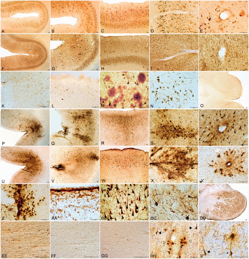
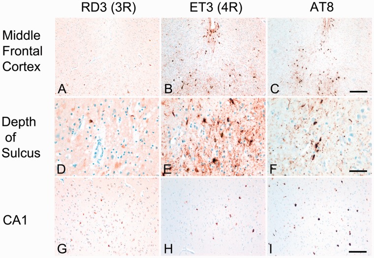
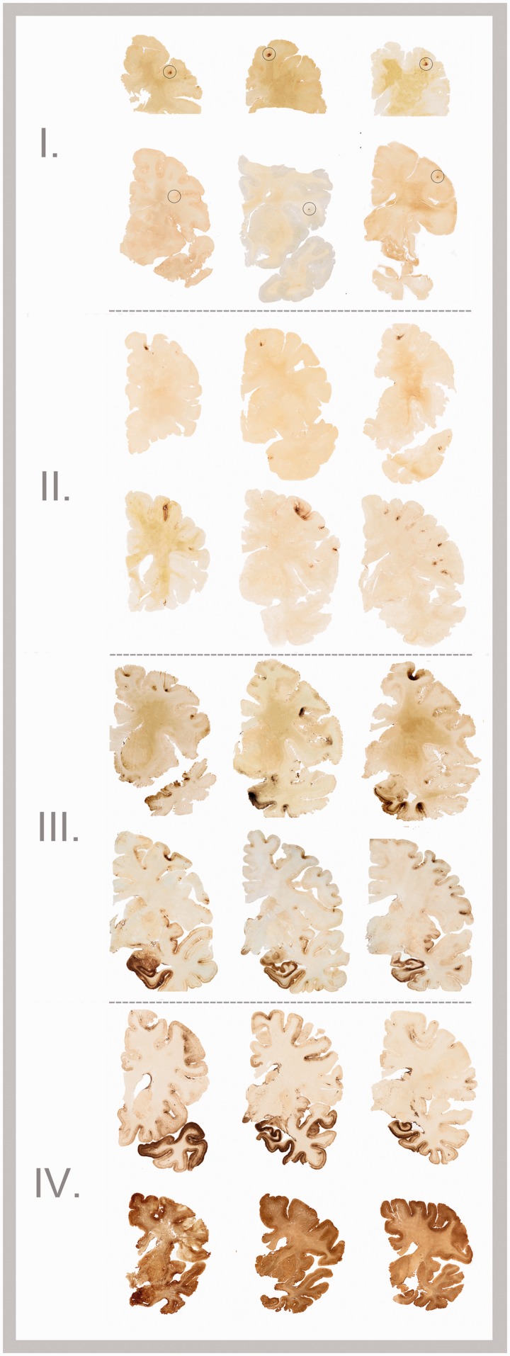
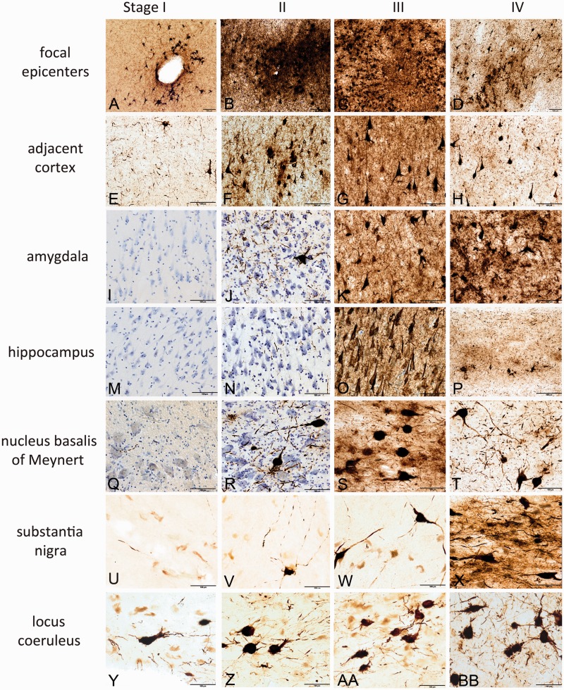
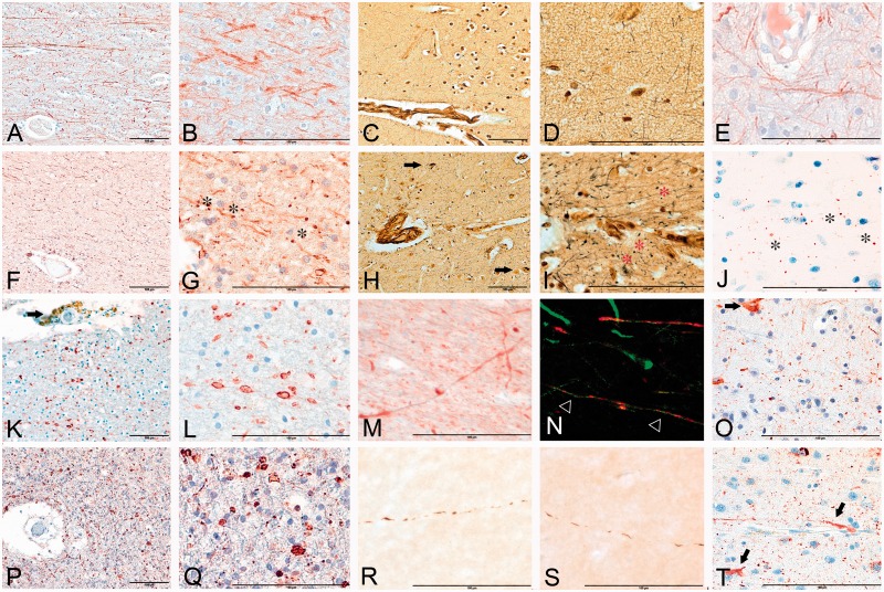
Chronic traumatic encephalopathy is a progressive tauopathy that occurs as a consequence of repetitive mild traumatic brain injury. We analysed post-mortem brains obtained from a cohort of 85 subjects with histories of repetitive mild traumatic brain injury and found evidence of chronic traumatic encephalopathy in 68 subjects: all males, ranging in age from 17 to 98 years (mean 59.5 years), including 64 athletes, 21 military veterans (86% of whom were also athletes) and one individual who engaged in self-injurious head banging behaviour. Eighteen age- and gender-matched individuals without a history of repetitive mild traumatic brain injury served as control subjects. In chronic traumatic encephalopathy, the spectrum of hyperphosphorylated tau pathology ranged in severity from focal perivascular epicentres of neurofibrillary tangles in the frontal neocortex to severe tauopathy affecting widespread brain regions, including the medial temporal lobe, thereby allowing a progressive staging of pathology from stages I-IV. Multifocal axonal varicosities and axonal loss were found in deep cortex and subcortical white matter at all stages of chronic traumatic encephalopathy. TAR DNA-binding protein 43 immunoreactive inclusions and neurites were also found in 85% of cases, ranging from focal pathology in stages I-III to widespread inclusions and neurites in stage IV. Symptoms in stage I chronic traumatic encephalopathy included headache and loss of attention and concentration. Additional symptoms in stage II included depression, explosivity and short-term memory loss. In stage III, executive dysfunction and cognitive impairment were found, and in stage IV, dementia, word-finding difficulty and aggression were characteristic. Data on athletic exposure were available for 34 American football players; the stage of chronic traumatic encephalopathy correlated with increased duration of football play, survival after football and age at death. Chronic traumatic encephalopathy was the sole diagnosis in 43 cases (63%); eight were also diagnosed with motor neuron disease (12%), seven with Alzheimer's disease (11%), 11 with Lewy body disease (16%) and four with frontotemporal lobar degeneration (6%). There is an ordered and predictable progression of hyperphosphorylated tau abnormalities through the nervous system in chronic traumatic encephalopathy that occurs in conjunction with widespread axonal disruption and loss. The frequent association of chronic traumatic encephalopathy with other neurodegenerative disorders suggests that repetitive brain trauma and hyperphosphorylated tau protein deposition promote the accumulation of other abnormally aggregated proteins including TAR DNA-binding protein 43, amyloid beta protein and alpha-synuclein.


Figures

Figure
1
Figure 1
Distinctive p-tau pathology of CTE compared with Alzheimer’s disease. (AO) Alzheimer’s disease. (AC) Double immunostained sections for amyloid-ß (red) and PHF-1 (brown) show diffuse cortical distribution of neurofibrillary tangles preferentially involving laminae III and V and without accentuation at depths of sulci. (D and E) Small blood vessels at sulcal depths show no clustering of neurofibrillary pathology perivascularly. (F and G) Sections immunostained for AT8 demonstrate diffuse cortical distribution of p-tau pathology without accumulations at sulcal depths. (H) Neurofibrillary tangles are preferentially distributed in laminae III and V. (I and J) Small blood vessels at bottom of cortical sulcus show no clustering of neurofibrillary pathology around vasculature (AT8 immunostain). (K) Subpial region at depth of sulcus shows no p-tau positive astrocytic tangles (AT8 immunostain). (L) Periventricular region of third ventricle shows no ependymal immunostaining for p-tau and low densities of p-tau neurites (AT8 immunostain). (M) Double immunostained section showing abundant amyloid-ß plaques (red) and interspersed PHF-1 neurofibrillary tangles (brown). (N) Moderate neurofibrillary change in substantia nigra pars compacta typical of severe Alzheimer’s disease (AT8 immunostain). (O) Absence of astrocytic tangles or neurofibrillary tangles in mammillary body in Alzheimer’s disease (AT8 immunostain). (PFF) CTE. (PR and UW) Sections immunostained for AT8 showing irregular cortical distribution of p-tau pathology with prominent subpial clusters of p-tau astrocytic tangles, focal accentuation at depths of sulci and distribution of neurofibrillary tangles in superficial cortical laminae II–III. (S, T, X and Y) Small blood vessels at bottom of cortical sulcus prominent perivascular distribution of astrocytic tangles and neurofibrillary tangles (AT8). (Z) Subpial region at depth of sulcus shows prominent cluster of AT8 positive astrocytic tangles. (AA) Periventricular region of third ventricle shows intense ependymal immunostaining for AT8 and abundant pericapillary neurites. (BB) Double immunostained section for amyloid-ß (red) and PHF-1 (brown) shows dense neurofibrillary tangles without amyloid-ß deposition. (CC) Dense AT8 immunostained astrocytic tangles and neurofibrillary tangles in substantia nigra pars compacta of severe CTE (AT8). (DD) Dense AT8 immunostained astrocytic tangles and neurofibrillary tangles in mammillary body typical of CTE. (EE) Dense CP-13 immunostained axonal varicosities and neuropil threads in the anterior commissure in CTE. (FF) AT8 immunostained axonal varicosities and neuropil threads in the external capsule in CTE. (GG) Alzheimer’s disease: low densities of AT8 immunostained neuropil threads in subcortical white matter. (HH and II) CTE: AT8 immunostained astrocytic tangles (open arrowheads), neurofibrillary tangles (arrowheads) and pre-tangles (asterisk) characteristic of CTE. Some sections counter-stained with cresyl violet; all scale bars = 100 µm.
Figure 2
Figure 2
Patterns of 3R and 4R Tau in CTE. (A, D and G) 3R immunostaining shows scattered RD3 immunoreactive neurons in middle frontal cortex and CA1 hippocampus. (B and E) 4R immunostaining shows many ET3 immunoreactive neurons and astrocytic tangles in the subpial region of the middle frontal cortex and at the depth of the sulcus. (H) 4R immunostaining shows many ET3 immunoreactive neurons in CA1 hippocampus. (C, F and I) AT8 immunostaining shows 3R and 4R immunopositive neurons and astrocytic tangles in middle frontal cortex and CA1 hippocampus. All are 10 -µm paraffin-embedded sections, scale bars = 50 µm.
Figure
3
Figure 3
The four stages of CTE. In stage I CTE, p-tau pathology is restricted to discrete foci in the cerebral cortex, most commonly in the superior, dorsolateral or lateral frontal cortices, and typically around small vessels at the depths of sulci (black circles). In stage II CTE, there are multiple epicentres at the depths of the cerebral sulci and localized spread of neurofibrillary pathology from these epicentres to the superficial layers of adjacent cortex. The medial temporal lobe is spared neurofibrillary p-tau pathology in stage II CTE. In stage III, p-tau pathology is widespread; the frontal, insular, temporal and parietal cortices show neurofibrillary degeneration with greatest severity in the frontal and temporal lobe, concentrated at the depths of the sulci. Also in stage III CTE, the amygdala, hippocampus and entorhinal cortex show neurofibrillary pathology. In stage IV CTE, there is severe p-tau pathology affecting most regions of the cerebral cortex and the medial temporal lobe, sparing calcarine cortex in all but the most severe cases. All images, CP-13 immunostained 50-µm tissue sections.
Figure 4
Figure 4
Hyperphosphorylated tau pathology in the four stages of CTE. In stage I CTE (first column), p-tau pathology is found in limited discrete perivascular foci (A), typically at the depths of sulci or around small vessels. There is mild p-tau pathology in cerebral cortices neighbouring the epicentres (E). There is no or minimal p-tau pathology in the amygdala (I) or CA1 of hippocampus (M). Occasional p-tau neurites are found in the nucleus basalis of Meynert (Q) and substantia nigra (U); isolated neurofibrillary tangles are present in the locus coeruleus (Y) in stage I. In stage II CTE (second column), there is spread of pathology from focal epicentres (B) to the superficial layers of adjacent cortex (F). The medial temporal lobe shows only mild neurofibrillary pathology, including amygdala (J) and CA1 hippocampus (N). Nucleus basalis of Meynert (R) and locus coeruleus (Z) demonstrate moderate p-tau pathology as neurofibrillary tangles and neurites; the substantia nigra (V) shows only modest pathology. In stage III, p-tau pathology is severe and widespread throughout the frontal, insular, temporal and parietal cortices. The cortical epicentres and depths of the sulci often consist of confluent masses of neurofibrillary tangles and astrocytic tangles (C). The intervening cortices show advanced neurofibrillary degeneration (G). The amygdala (K), hippocampus (O) and entorhinal cortex demonstrate marked neurofibrillary pathology. The nucleus basalis of Meynert shows dense neurofibrillary tangles (S); the locus coeruleus (AA) shows advanced neurofibrillary pathology, and the substantia nigra is moderately affected (W) in stage III CTE. In stage IV CTE, there is widespread p-tau pathology affecting most regions of the cerebral cortex and medial temporal lobe with relative sparing of the calcarine cortex. Astrocytic tangles are prominent, and there is marked neuronal loss in the cortex, amygdala and hippocampus. Phosphorylated-tau neurofibrillary tangles are reduced in size and density. The cortical epicentres show severe neuronal loss and prominent astrocytic tangles (D); similar changes are found throughout the frontal, temporal and parietal cortices (H). The amygdala demonstrates intense gliosis and p-tau neuronal and glial degeneration (L). The hippocampus is sclerotic with marked neuronal loss, gliosis, ghost neurofibrillary tangles and astrocytic tangles (P). The nucleus basalis of Meynert shows marked neurofibrillary pathology and gliosis (T); the substantia nigra (X) and locus coeruleus (BB) show advanced neurofibrillary pathology. All images: CP-13 immunostained 50 -µm tissue sections, some counterstained with cresyl violet, all scale bars = 100 µm.
Figure 5
Figure 5
The axonal pathology of CTE. (A and B) Phosphorylated neurofilament immunostaining for SMI-34 (red) in control subcortical white matter shows regular alignment and linear morphology of most axonal profiles. (C and D) Axons in control cerebral cortex stained with Bielschowsky’s silver method show a fine linear pattern and regularity. (E) Phosphorylated neurofilament immunostaining (SMI-34) in control cerebral cortex also shows regular alignment and linear morphology of axons even around small vessels. (F and G) Phosphorylated neurofilament immunostaining in cerebral cortex of stage II CTE demonstrates alterations in alignment and numerous rounded axonal varicosities around small vessels (asterisks). (H and I) Bielschowsky silver method in stage I CTE shows neurofibrillary tangles (arrows) and silver positive axonal varicosities (asterisks) around small arteriole. (J) SMI-34 immunostaining of same cortical focus as in H and I also shows axonal varicosities (asterisks) around cortical arteriole. (K and L) SMI-34 immunostaining in cerebral cortex of stage III CTE shows marked reduction in axonal staining and numerous large, irregular axonal varicosities. A small arteriole shows marked infiltration with haemosiderin-laden macrophages (arrow). (M) Axonal varicosities and irregularities are also found in stage I CTE. (N) CTE stage II double immunostained for phosphorylated tau (PHF-1, brown) and phosphorylated neurofilament (SMI-34, red) shows axonal swellings in continuity with phosphorylated tau neuritic abnormalities. (O) Double immunofluorescence staining for phosphorylated neurofilament, SMI-34 (red) and PHF-1 (green) in the subcortical white matter of CTE stage III demonstrates contiguous axonal varicosities (red) as well as p-tau (green) (arrowheads) in the axon. (P and Q) SMI-34 immunostaining in subcortical white matter of stage IV CTE shows severe axonal loss and multiple large, irregular axonal varicosities. (R and S) Phosphorylated tau (AT8, brown) immunoreactive irregular axonal profiles found in deep white matter tracts CTE stage I. (T) Dense axonal varicosities, distorted axonal profiles and neurofibrillary tangles (arrows) characterize the cerebral cortex of CTE stage IV immunostained with phosphorylated neurofilament (SMI-34). Images from 10 µm tissue sections, scale bars = 100 µm, except O = 10 µm.
Figure 6
Figure 6
The phosphorylated TDP43 pathology of CTE and CTE-MND. (A) pTDP-43 immunostained neurites found in periventricular region of the third ventricle in CTE II. (B) CTE stage II demonstrating pTDP-43 immunostained neurites in a perivascular distribution. (C) pTDP-43 immunoreactive inclusion in locus coeruleus in CTE stage II. (D) Clusters of pTDP-43 immunoreactive neurites in the subpial region of the brainstem in stage III CTE. (E) Perivascular pTDP-43 neurites in stage III CTE. (F) Subpial pTDP-43 neurites in the corpus callosum of stage IV CTE. (G) Dense pTDP-43 abnormalities in the temporal cortex of stage IV CTE-MND. (H) Dense pTDP-43 pathology of CA1 hippocampus in stage IV CTE. (IO) (I) perivascular focus at sulcal depth pTDP-43 abnormalities are widespread throughout the CNS in CTE associated with MND (CTE-MND) (J, corpus callosum; K, cerebral peduncle; L, fornix; M, subpial region of frontal cortex; N, perivascular region frontal cortex; O, Rolandic cortex). All images: 50-µm tissue sections, all scale bars = 100 µm.

References

    1. Amadoro G, Ciotti MT, Costanzi M, Cestari V, Calissano P, Canu N. NMDA receptor mediates tau-induced neurotoxicity by calpain and ERK/MAPK activation. Proc Natl Acad Sci USA. 2006;103:2892–7. - PMC - PubMed
    1. Anthony IC, Norrby KE, Dingwall T, Carnie FW, Millar T, Arango JC, et al. Predisposition to accelerated Alzheimer-related changes in the brains of human immunodeficiency virus negative opiate abusers. Brain. 2010;133:3685–98. - PubMed
    1. Arriagada PV, Growdon JH, Hedley-Whyte ET, Hyman BT. Neurofibrillary tangles but not senile plaques parallel duration and severity of Alzheimer’s disease. Neurology. 1992;42:631–9. - PubMed
    1. Baugh CM, Stamm JM, Riley DO, Gavett BE, Shenton ME, Lin A, et al. Chronic traumatic encephalopathy: neurodegeneration following repetitive concussive and subconcussive brain trauma. Brain Imaging Behav. 2012;2:244–54. - PubMed
    1. Bigio EH. Update on recent molecular and genetic advances in FTLD. J Neuropathol Exp Neurol. 2008;67:635–48. - PMC - PubMed

Publication types