

Review
doi: 10.1002/wsbm.1201.
Epub 2012 Dec 3.
Lymphatic vessels in health and disease
Affiliations
- PMID: 23209022
- PMCID: PMC3527689
- DOI: 10.1002/wsbm.1201
Item in Clipboard
Review
Lymphatic vessels in health and disease
Wiley Interdiscip Rev Syst Biol Med.
2013 Jan-Feb.
Abstract
The lymphatic vasculature plays vital roles in tissue fluid balance, immune defense, metabolism, and cancer metastasis. In adults, lymphatic vessel formation and remodeling occur primarily during inflammation, development of the corpus luteum, wound healing, and tumor growth. Unlike the blood circulation, where unidirectional flow is sustained by the pumping actions of the heart, pumping actions intrinsic to the lymphatic vessels themselves are important drivers of lymphatic flow. This review summarizes critical components that control lymphatic physiology.
Copyright © 2012 Wiley Periodicals, Inc.
Conflict of interest statement
Figures
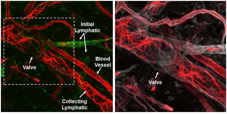
The lymphatic vessels of the ear of an athymic nude mouse are shown. LYVE-1 (green) indicates the initial lymphatic vessels. αSMA (red) indicates the SMCs of the collecting lymphatic vessels and blood vessels. The circumferential αSMA staining pattern of the collecting lymphatic vessels is distinct from the more homogenous pattern of the blood vessels. CD31 (white) indicates all endothelial cells in the field and shows an intraluminal valve in the collecting lymphatic vessel.
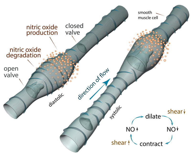
This illustration of the diastolic and systolic phases of an autonomous lymphatic contraction shows the NO dependency. In the diastolic phase, local NO release allows for the relaxation of the vessel wall and filling to occur. As the NO degrades, the vessel constricts, driving flow into the next lymphangion. It is hypothesized that the increase in flow and shear stress as a result of a contraction, stimulates NO production, allowing the diastolic filling to occur. The spatial and temporal gradients of NO are critical to proper contraction function and are mediated by eNOS in LECs.
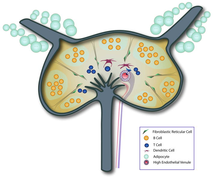
This simplified schematic of the lymph node highlights key structural features critical for the proper activation of an immune response. The adipose-encased afferent collecting lymphatic vessels move antigen-rich lymph into the subcapsular sinus. Fluid and small antigens can then filter into the lymph node cortex, where B cell follicles are found. Reticular fibers, bound by their associated FRCs and specialized DCs, traverse the cortex to rapidly bring antigen to the paracortical and medullary regions where T cells reside. HEVs in the paracortical area bring naïve T cells into the node as well to interact with DCs. In the medulla, there are lymphatic vessels that drain the lymph node and collect fluid into the efferent lymphatic vessel.
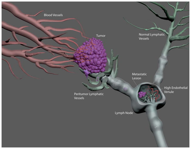
In contrast to functional blood vessels that can be found throughout the tumor, functional lymphatic vessels are found in the margin of tumors. These tumor margin lymphatic vessels tend to be enlarged and have greater lymph flow compared to lymphatic vessels draining normal tissues. These functional lymphatics are penetrated by invading cancer cells, which travel to the draining lymph node where they evade the immune system and start to form a secondary metastatic tumor. Understanding the growth of the cancer cells in the lymph node is critical to the development of effective treatment for these metastatic lesions.
References
-
- Sebzda E, Hibbard C, Sweeney S, Abtahian F, Bezman N, Clemens G, Maltzman JS, Cheng L, Liu F, Turner M, et al. Syk and Slp-76 mutant mice reveal a cell-autonomous hematopoietic cell contribution to vascular development. Dev Cell. 2006;11:349–361. - PubMed
-
- Wigle JT, Oliver G. Prox1 function is required for the development of the murine lymphatic system. Cell. 1999;98:769–778. - PubMed
Publication types
MeSH terms
Grants and funding
- T32 CA073479/CA/NCI NIH HHS/United States
- R00CA137167/CA/NCI NIH HHS/United States
- K99 HL111343/HL/NHLBI NIH HHS/United States
- R21 AI097745/AI/NIAID NIH HHS/United States
- DP2OD008780/OD/NIH HHS/United States
- T32CA073479/CA/NCI NIH HHS/United States
- R00 CA137167/CA/NCI NIH HHS/United States
- R01 CA149285/CA/NCI NIH HHS/United States
- R01CA149285/CA/NCI NIH HHS/United States
- R01 HL106584/HL/NHLBI NIH HHS/United States
- R21AI097745/AI/NIAID NIH HHS/United States
- R01HL106584/HL/NHLBI NIH HHS/United States
- P01 CA080124/CA/NCI NIH HHS/United States
- DP2 OD008780/OD/NIH HHS/United States
LinkOut - more resources
Full Text Sources
Other Literature Sources
Medical
