Review
. 2013 Feb 1;5(2):a008029.
doi: 10.1101/cshperspect.a008029.

Wnt signaling in skin development, homeostasis, and disease

Affiliations
Review

Wnt signaling in skin development, homeostasis, and disease

Xinhong Lim et al. Cold Spring Harb Perspect Biol. .

Abstract

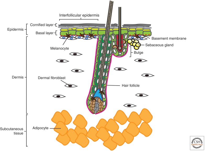
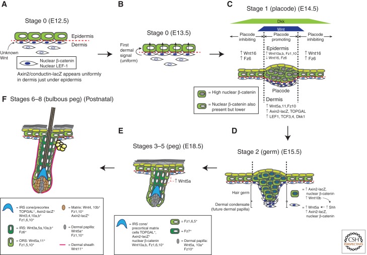
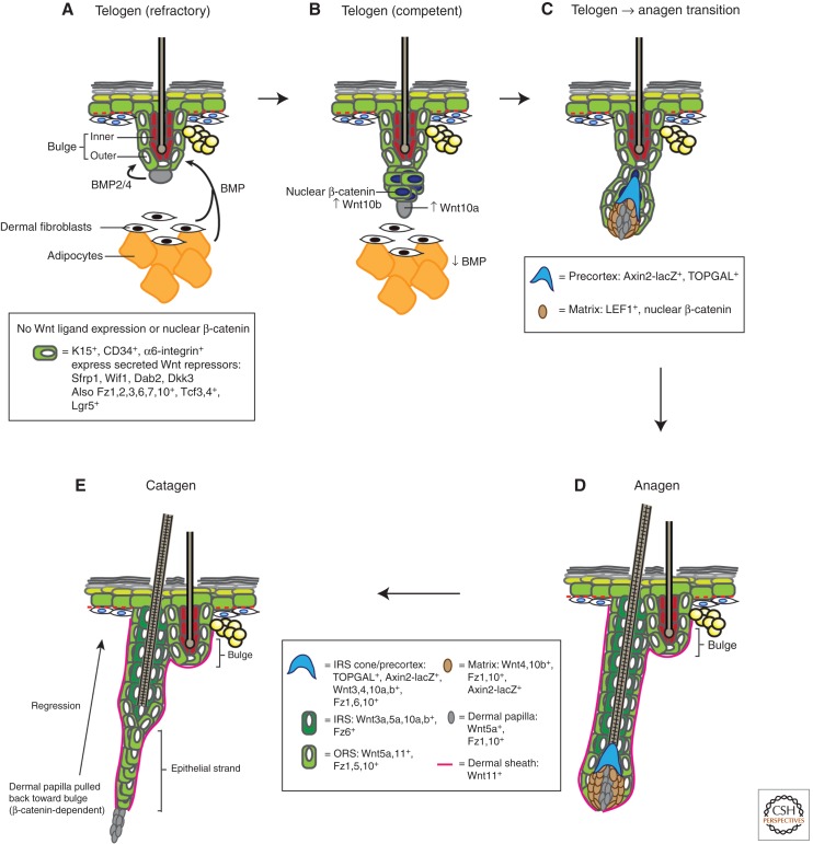
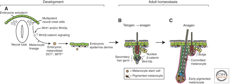
The skin and its appendages constitute the largest organ of the body. Its stratified epithelia offer protection from environmental stresses such as dehydration, irradiation, mechanical trauma, and pathogenic infection, whereas its appendages, like hair and sebaceous glands, help regulate body temperature as well as influence animal interaction and social behavior through camouflage and sexual signaling. To respond to and function effectively in a dynamic external environment, the skin and its appendages possess a remarkable ability to regenerate in a carefully controlled fashion. When this finely tuned homeostatic process is disrupted, skin diseases such as cancers may result. At present, the molecular signals that orchestrate cell proliferation, differentiation, and patterning in the skin remain incompletely understood. It is increasingly apparent that many morphogenetic pathways with key roles in development are also important in regulating skin biology. Of these, Wnt signaling has emerged as the dominant pathway controlling the patterning of skin and influencing the decisions of embryonic and adult stem cells to adopt the various cell lineages of the skin and its appendages, as well as subsequently controlling the function of differentiated skin cells. Here we will review established concepts and present recent advances in our understanding of the diverse roles that Wnt signaling plays in skin development, homeostasis, and disease.


Figures

Figure 1.
Figure 1.
Anatomy of mammalian skin. The skin is a complex composite of multiple miniorgans. The topmost layer is a stratified epidermis consisting of keratinocytes that differentiate as they progress from the basal layer to the suprabasal and eventually form dead enucleated squames that are sloughed off from the cornified layer. Multiple different appendages branch from the epidermal layer, including hair follicles (HFs), sebaceous glands, and sweat glands. The dermis, which lies below the epidermis and surrounds the HFs, is composed of fibroblasts of mesenchymal origin that reside in a fibrous matrix that they produce. Melanocytes are interspersed among the keratinocytes of the epidermis and HF (bulge and matrix), where they pigment the skin and hair. Below all these is a subcutaneous layer of adipocytes that serve to insulate the animal.
Figure 2.
Figure 2.
Wnt signaling during skin development and hair follicle morphogenesis. Hair follicle (HF) morphogenesis proceeds in three phases: induction (AC), organogenesis (D,E), and cytodifferentiation (F), which can be further separated into eight stages (for a detailed review of these stages see Paus et al. 1999 and Schneider et al. 2009). The morphology and molecular events occurring at each stage are depicted. Data for developmental timings are from Blanpain and Fuchs (2006) and Zhang et al. (2009). Expression data are from DasGupta and Fuchs (1999), Reddy et al. (2001, 2004), and Zhang et al. (2009). Induction: (A) Stage 0 (E12.5): Before any visible hair placode formation, dermal fibroblasts appear to uniformly receive an unidentified Wnt signal from an unknown source, resulting in nuclear localization of β-catenin. (B) Stage 0 (E13.5): Dermal fibroblasts then produce the first dermal signal, which is likely also a Wnt. (C) Stage 1 (E14.5): On receiving the first dermal signal, Wnt/β-catenin signaling becomes active in the epidermis and is especially elevated at placode-forming regions. Reaction–diffusion reactions between slower-diffusing activating Wnts and faster-diffusing inhibitory Dkks produce a regular array of these high-Wnt placode-forming regions that eventually result in patterned hair growth (see also Box 1). The nascent epidermal placode cells proliferate and accumulate and also start producing Wnt ligands like Wnt10b. Organogenesis: (D) Stage 2 (E15.5): As the placode develops into a visible hair germ, the Wnt ligands it produces induce underlying fibroblasts to form a dermal condensate that will eventually develop into a follicular dermal papilla. This process is also dependent on Shh pathway activity in the dermal condensates. (E) Stages 3–5 (E18.5): As the hair germ grows into a bulbous hair peg, differentiating epithelial layers become apparent. Wnt/β-catenin is evident in these layers. Cytodifferentiation: (F) Stages 6–8 (bulbous peg) (early postnatal): The morphology and biochemistry of the growing HF resembles that of a postnatal anagen follicle. Wnt/β-catenin signaling also seems to be involved in determining the specific follicular lineages that progenitor keratinocytes adopt, with strong and complex expression of Wnt ligands, receptors, and pathway reporters in the various follicular layers.
Figure 3.
Figure 3.
Wnt signaling during hair follicle cycling. Postnatal hair follicles (HFs) cycle repeatedly through rest (telogen), growth (anagen), and death/regression (catagen). The morphology and molecular events taking place at each stage of the hair cycle are depicted (for a comprehensive histological review of hair cycle stages see Müller-Röver et al. 2001). Expression data are from DasGupta and Fuchs (1999), Reddy et al. (2001, 2004), Blanpain et al. (2004), Morris et al. (2004), Tumbar et al. (2004), and Zhang et al. (2009). (A) During refractory telogen (BMP-high, Wnt-low) (Plikus et al. 2008), BMP is secreted from adipocytes and dermal fibroblasts, as well as the dermal papilla, to maintain the HF bulge in a low-growth, quiescent state. The bulge is thought to also be in a Wnt-inhibited state, by expressing various Wnt pathway inhibitors. (B) In the competent telogen phase (BMP-low, Wnt-high), BMP expression in adipocytes and dermal cells is reduced, allowing bulge cells to respond to anagen-inducing cues. One such cue is likely to involve an as-yet-unidentified Wnt. At this time, Wnt/β-catenin signaling becomes up-regulated in bulge and secondary hair germ cells, and they produce Wnt ligands such as Wnt10b. (C) As the follicle transitions from telogen to anagen and starts growing, bulge/secondary hair germ keratinocytes proliferate and migrate along the developing outer root sheath (ORS) to enwrap the dermal papilla and push it downward, away from the bulge. Wnt/β-catenin signaling continues to be active in these keratinocytes that form the developing matrix and the precortex. (D) The HF continues to grow downward in anagen. Distinct, differentiated layers (sheaths) are observed, each expressing different Wnt ligands and receptors. The significance of these complex expression patterns remains unknown. Lgr5+ follicle stem cells are found throughout the ORS and likely migrate to the matrix, where they differentiate into keratinocytes populating the various lineages of the anagen HF. Wnt/β-catenin signaling is also active in the matrix and is thought to be important for specifying these lineages. It is unclear if particular Wnts are responsible for specifying particular anagen lineages. (E) As the follicle exits anagen and enters catagen, it stops growing and starts dying. The follicle regresses and forms an epithelial strand that slowly pulls the dermal papilla upward to the level of the bulge, a process that is dependent on β-catenin (van Genderen et al. 1994).
Figure 4.
Figure 4.
Wnt signaling during melanocyte development and homeostasis. (A) As the embryo develops, multipotent neural crest cells receive Wnt signals that instruct them to adopt the melanocyte lineage and form melanoblasts. These melanoblasts migrate and populate the embryonic epidermis and developing hair follicles (HFs). (B) Embryonic melanoblasts eventually give rise to adult melanocyte stem cells, which reside in the telogen bulge in close proximity to follicular stem cells. Follicular and melanocyte stem cells regulate each other and coordinately activate Wnt/β-catenin signaling at the onset of anagen (Rabbani et al. 2011). (C) The activation of Wnt signaling in melanocyte stem cells causes them to proliferate and differentiate into melanocytes, which then migrate along the outer root sheath (ORS) to enter the matrix. In the matrix, they potentially receive additional Wnt signals that cause them to transfer pigment to differentiating cortical keratinocytes, producing a pigmented hair shaft.

References

    1. Adaimy L, Chouery E, Megarbane H, Mroueh S, Delague V, Nicolas E, Belguith H, de Mazancourt P, Megarbane A 2007. Mutation in WNT10A is associated with an autosomal recessive ectodermal dysplasia: The odonto-onycho-dermal dysplasia. Am J Hum Genet 81: 821–828 - PMC - PubMed
    1. Ambler CA, Määttä A 2009. Epidermal stem cells: Location, potential and contribution to cancer. J Pathol 217: 206–216 - PubMed
    1. Andl T, Reddy ST, Gaddapara T, Millar SE 2002. WNT signals are required for the initiation of hair follicle development. Dev Cell 2: 643–653 - PubMed
    1. Andl T, Ahn K, Kairo A, Chu EY, Wine-Lee L, Reddy ST, Croft NJ, Cebra-Thomas JA, Metzger D, Chambon P, et al. 2004. Epithelial Bmpr1a regulates differentiation and proliferation in postnatal hair follicles and is essential for tooth development. Development 131: 2257–2268 - PubMed
    1. Atit R, Sgaier SK, Mohamed OA, Taketo MM, Dufort D, Joyner AL, Niswander L, Conlon RA 2006. β-Catenin activation is necessary and sufficient to specify the dorsal dermal fate in the mouse. Dev Biol 296: 164–176 - PubMed

Publication types

LinkOut - more resources