. 2013 Jan 11;288(2):1088-98.
doi: 10.1074/jbc.M112.393090. Epub 2012 Dec 3.

The developing chicken yolk sac acquires nutrient transport competence by an orchestrated differentiation process of its endodermal epithelial cells

Affiliations

The developing chicken yolk sac acquires nutrient transport competence by an orchestrated differentiation process of its endodermal epithelial cells

Raimund Bauer et al. J Biol Chem. .

Abstract

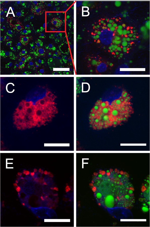
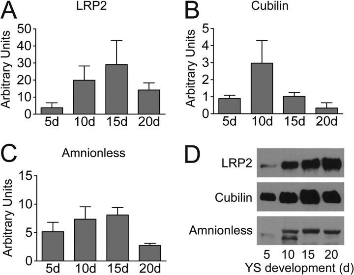
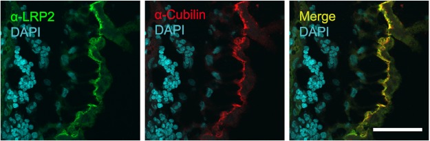
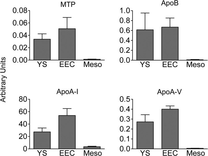
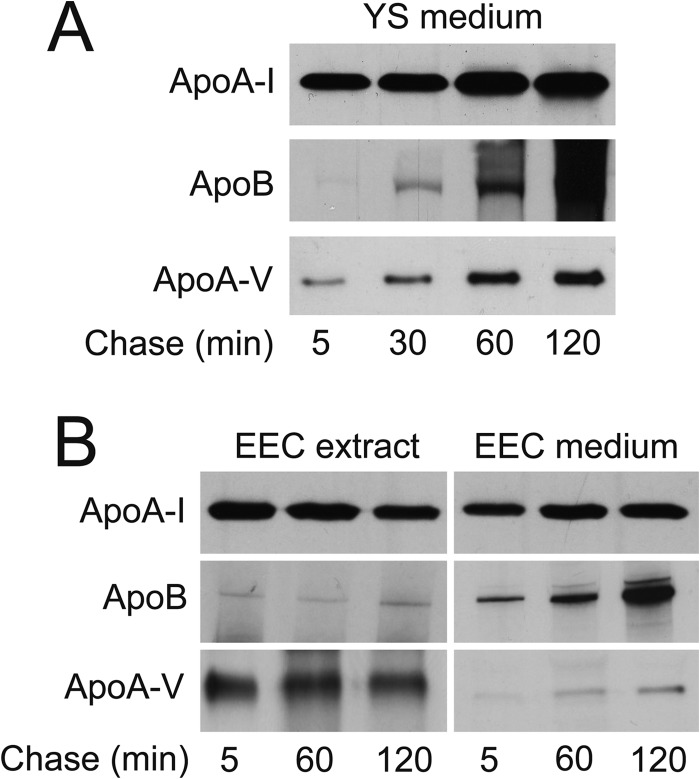
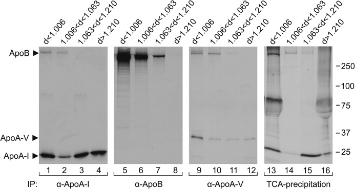
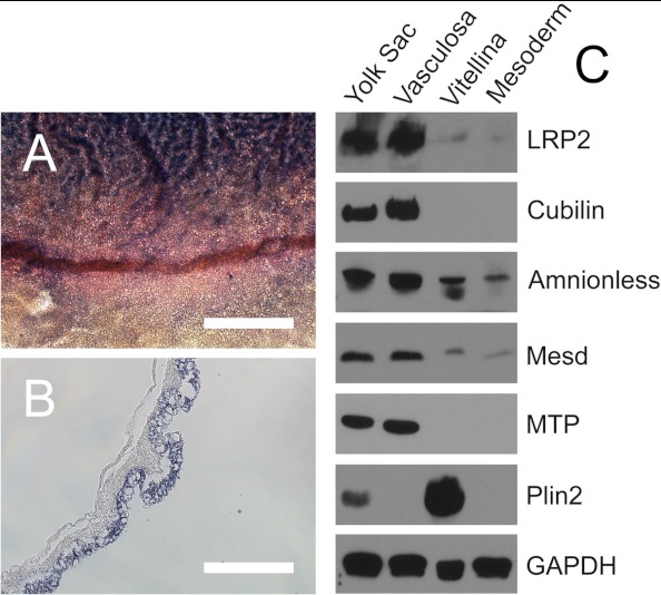
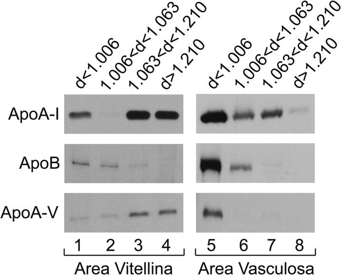
During chicken yolk sac (YS) growth, mesodermal cells in the area vasculosa follow the migrating endodermal epithelial cell (EEC) layer in the area vitellina. Ultimately, these cells form the vascularized YS that functions in nutrient transfer to the embryo. How and when EECs, with their apical aspect directly contacting the oocytic yolk, acquire the ability to take up yolk macromolecules during the vitellina-to-vasculosa transition has not been investigated. In addressing these questions, we found that with progressive vascularization, the expression level in EECs of the nutrient receptor triad, LRP2-cubilin-amnionless, changes significantly. The receptor complex, competent for uptake of yolk proteins, is produced by EECs in the area vasculosa but not in the area vitellina. Yolk components endocytosed by LRP2-cubilin-amnionless, preformed and newly formed lipid droplets, and yolk-derived very low density lipoprotein, shown to be efficiently endocytosed and lysosomally processed by EECs, probably provide substrates for resynthesis and secretion of nutrients, such as lipoproteins. In fact, as directly demonstrated by pulse-chase experiments, EECs in the vascularized, but not in the avascular, region efficiently produce and secrete lipoproteins containing apolipoprotein A-I (apoA-I), apoB, and/or apoA-V. In contrast, perilipin 2, a lipid droplet-stabilizing protein, is produced exclusively by the EECs of the area vitellina. These data suggest a differentiation process that orchestrates the vascularization of the developing YS with the induction of yolk uptake and lipoprotein secretion by EECs to ensure embryo nutrition.


Figures

FIGURE 1.
FIGURE 1.
Endodermal epithelial cells take up yVLDL particles. Explants from YS were transferred to Lab Tek Permanox 4-well imaging chambers. After 48 h of EEC outgrowth, the medium was changed to serum-free conditions, and 50 μg/ml DiI-labeled yVLDL (red fluorescence) was added. In A and B, the EECs were incubated with the labeled lipoprotein for 4 h at 37 °C; B shows the magnification of a cell from A. In C–F, the cells were incubated with 50 μg/ml DiI-yVLDL for 4 h, the medium was removed and replaced with medium without DiI-yVLDL, and cells were further incubated for 20 h. In E and F, the incubation medium contained 200 μm chloroquine, an inhibitor of lysosomal degradation. After the incubations, the cells were washed two times with PBS, fixed in 4% PFA in PBS, pH 7.4, for 30 min at room temperature, and washed two times with PBS. After counterstaining with BODIPY 493/503 (Molecular Probes) for the visualization of lipid droplets (green fluorescence) and DAPI for the nuclei, respectively, cells were mounted in Fluorescent Mounting Medium (Dako). Images were obtained with a Zeiss LSM510 confocal microscope. Scale bar, 50 μm (A) or 20 μm (B–F).
FIGURE 2.
FIGURE 2.
Expression of LRP2, cubilin, and amnionless in the chicken yolk sac at 5, 10, 15, and 20 days of development. mRNA levels in the yolk sac of LRP2 (A), cubilin (B), and amnionless (C) were determined using qPCR as described under “Experimental Procedures.” Transcript levels are expressed as arbitrary units relative to chicken β-actin and represent the average of at least triplicate measurements ± S.E. (error bars). D, analysis of LRP2, cubilin, and amnionless protein levels by Western blotting. Fifty micrograms of membrane protein per lane from yolk sacs of the indicated developmental stages were separated either by 6% (LRP2 and cubilin) or 12% (amnionless) SDS-PAGE under non-reducing conditions, blotted to nitrocellulose membranes, and probed with antisera directed against ggLRP2, ggCubilin, and ggAmnionless, followed by an HRP-coupled anti-rabbit secondary antibody as described under “Experimental Procedures.”
FIGURE 3.
FIGURE 3.
The multiligand receptors LRP2 and cubilin colocalize at the apical surface of the endodermal epithelial cells of the yolk sac. Cryosections (4 μm) of the area vasculosa of yolk sac tissue from 10-day-old chicken embryos were prepared as described under “Experimental Procedures.” The sections were stained with our rabbit anti-ggLRP2 antiserum (green staining) and our mouse anti-ggCubilin antiserum (red staining). The merged image is shown on the right. For the visualization of cell nuclei, the sections were counterstained with DAPI. Images were acquired using a Zeiss LSM 510 confocal microscope. Scale bar, 50 μm.
FIGURE 4.
FIGURE 4.
The yolk sac endodermal epithelial cells, but not mesodermal cells, express genes for lipoprotein assembly. Transcript levels of apoA-I, apoB, apoA-V, and MTP were analyzed with qPCR in the YS and, after dissection, in the EECs and in cells of the mesodermal layer (Meso). Transcript levels are expressed as arbitrary units relative to chicken β-actin and represent the average of at least triplicate measurements ± S.E. (error bars).
FIGURE 5.
FIGURE 5.
Endodermal epithelial cells of the yolk sac synthesize and secrete apolipoproteins. Whole yolk sac tissue explants (A) and EECs from the area vasculosa (B) were pulse-labeled with [35S]methionine/cysteine for 1 h and chased for the indicated time periods. Radiolabeled secreted apolipoproteins were immunoprecipitated with specific antisera/antibodies directed against apoA-I, apoB, and apoA-V from the conditioned media (A and B) and cellular apolipoproteins from cell extracts (B) as described under “Experimental Procedures.”
FIGURE 6.
FIGURE 6.
Distribution of apolipoproteins A-I, B, and A-V among yolk sac-derived lipoprotein fractions. 35S-Labeled lipoproteins secreted by YS tissue were isolated and fractionated as described under “Experimental Procedures” and subsequently were immunoprecipitated (IP) with the indicated respective antisera/antibodies under detergent-free conditions. Equal amounts of isolated lipoprotein fractions were precipitated with TCA as described under “Experimental Procedures.” Autoradiography was performed after 4–18% SDS-PAGE of the precipitates.
FIGURE 7.
FIGURE 7.
EECs in the area vasculosa and area vitellina display opposite levels of proteins involved in yolk uptake and resynthesis of lipoproteins. Whole-mount in situ hybridization was performed with YS tissue at day 5 of embryonic development using digoxigenin-labeled antisense probes for chicken cubilin. A shows the transition zone from the area vasculosa (top) to the area vitellina (bottom). B, a YS section through the cubilin-positive area vasculosa. Scale bar, 800 μm in A and 200 μm in B. In C, fresh tissue samples of either the whole YS, the EECs of the area vasculosa (Vasculosa), the EECs of the area vitellina (Vitellina), or the blood vessel-containing mesodermal cell layer (Mesoderm) from 5-day chicken embryos were isolated, and total protein fractions were prepared as described under “Experimental Procedures.” Fifty micrograms of total protein lysate per lane from the indicated sources were separated either by 6% (LRP2 and cubilin) or 12% (amnionless, Mesd, MTP, Plin2, and GAPDH for loading control) SDS-PAGE under non-reducing conditions, blotted to nitrocellulose membranes, and probed with the indicated antisera/antibodies. For visualization of bound primary antibodies, corresponding HRP-coupled secondary antibodies were used as described under “Experimental Procedures.”
FIGURE 8.
FIGURE 8.
EECs in the area vitellina and in the area vasculosa show different apolipoprotein secretion patterns. Biosynthetically radiolabeled lipoproteins, secreted into the media of EEC explants from either the area vitellina or the area vasculosa, were isolated and fractionated as described under “Experimental Procedures” and immunoprecipitated with specific antisera/antibodies directed against apoA-I, apoB, or apoA-V. Autoradiography was performed after separation of the immunoprecipitated proteins by SDS-PAGE as described under “Experimental Procedures.” Exposure times for apoA-I immunoprecipitates were 16 h for lipoproteins derived from both YS areas. For apoB and apoA-V immunoprecipitates, exposure times were 21 days for both apolipoproteins derived from the area vitellina and 16 h and 10 days, respectively, for lipoprotein fractions derived from the area vasculosa.
FIGURE 9.
FIGURE 9.
The developing chicken YS: acquisition of nutrient transport function by differentiation of the EECs is linked to induction of vascularization. Three regions of the growing YS can be distinguished, each schematically represented by an EEC and associated cells. The three regions are the area vitellina (left), a transition zone (center), and the area vasculosa (right). The area vitellina EECs lack endocytic receptors and nonspecifically phagocytose yolk components while they migrate along the yolk surface underneath the ectoderm. Due to the presence of Plin2 on the area vitellina LD surface (red circle around LDs in area vitellina), the lipids of the LDs, particularly triglycerides, are not available for extensive lipidation of apoA-I, and therefore the predominant secreted lipoprotein particles have a high density. In the transition zone, characterized by migration of mesenchymal progenitor cells into the interstitium between ecto- and endoderm, receptors begin to be produced, phagocytotic yolk uptake continues, and as Plin2 levels drop, some LD lipids become available for increased lipoprotein synthesis and secretion. In the area vasculosa, blood vessel formation is coordinated with the enhanced production and localization of endocytic receptors with specificity for selected yolk components to the apical aspect of EECs, the disappearance of Plin2, and the expression of genes specifying MTP and additional apolipoproteins. The LDs are lipolyzed, and the lipids liberated from LDs plus components derived from uptake and lysosomal processing of lipoproteins, such as yVLDL, and of other yolk macromolecules are utilized for the assembly of lipoproteins containing newly synthesized apoA-I, apoB, and apoA-V (i.e. VLDL-like and HDL-like particles that become efficiently secreted for transfer into the adjacent blood vessels, thereby supplying the embryo with nutrients generated by transformation of oocytic yolk in the EECs).

References

    1. Zohn I. E., Sarkar A. A. (2010) The visceral yolk sac endoderm provides for absorption of nutrients to the embryo during neurulation. Birth Defects Res. A Clin. Mol. Teratol. 88, 593–600 - PubMed
    1. Schneider W. J., Osanger A., Waclawek M., Nimpf J. (1998) Oocyte growth in the chicken. Receptors and more. Biol. Chem. 379, 965–971 - PubMed
    1. Palis J., McGrath K. E., Kingsley P. D. (1995) Initiation of hematopoiesis and vasculogenesis in murine yolk sac explants. Blood 86, 156–163 - PubMed
    1. Stern C. D., Downs K. M. (2012) The hypoblast (visceral endoderm). An evo-devo perspective. Development 139, 1059–1069 - PMC - PubMed
    1. Yoshizaki N., Soga M., Ito Y., Mao K. M., Sultana F., Yonezawa S. (2004) Two-step consumption of yolk granules during the development of quail embryos. Dev. Growth Differ. 46, 229–238 - PubMed

Publication types