Review
. 2013 Jan 18;288(3):1806-13.
doi: 10.1074/jbc.R112.421909. Epub 2012 Dec 3.

Using hydrogen/deuterium exchange mass spectrometry to define the specific interactions of the phospholipase A2 superfamily with lipid substrates, inhibitors, and membranes

Affiliations
Review

Using hydrogen/deuterium exchange mass spectrometry to define the specific interactions of the phospholipase A2 superfamily with lipid substrates, inhibitors, and membranes

Jian Cao et al. J Biol Chem. .

Abstract

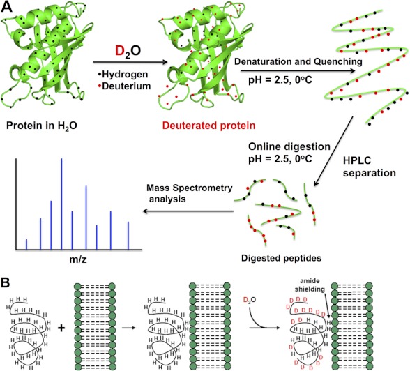
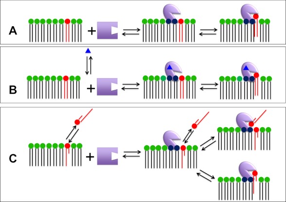
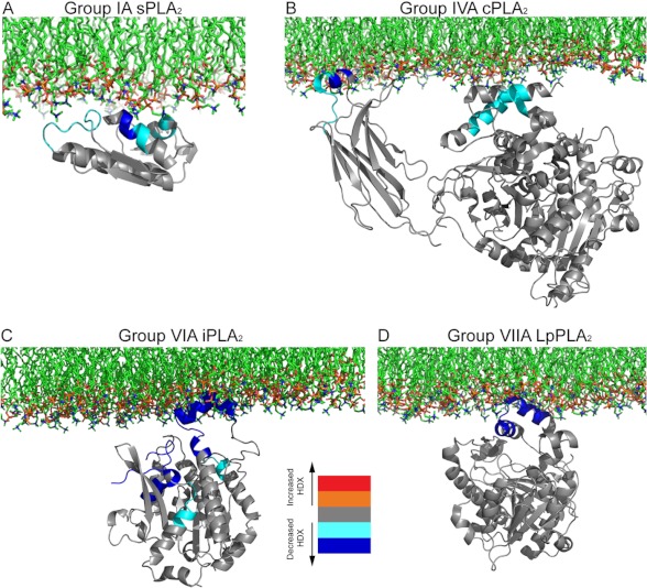
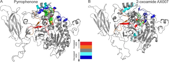
The phospholipase A(2) (PLA(2)) superfamily consists of 16 groups and many subgroups and constitutes a diverse set of enzymes that have a common catalytic activity due to convergent evolution. However, different PLA(2) types have unique three-dimensional structures and catalytic residues as well as specific tissue localization and distinct biological functions. Understanding how the different PLA(2) enzymes associate with phospholipid membranes, specific phospholipid substrate molecules, and inhibitors on a molecular basis has advanced in recent years due to the introduction of hydrogen/deuterium exchange mass spectrometry. Its theory, practical considerations, and application to understanding PLA(2)/membrane interactions are addressed.


Figures

FIGURE 1.
FIGURE 1.
Basis of H/D exchange. A, the protein of interest is incubated with deuterated water and labeled via exchangeable hydrogens. The exchange is then quenched to lock the deuterium atoms in position by lowering the pH of the protein-containing solution (which also denatures the protein) and lowering the solution temperature. The protein is then loaded onto an HPLC column that contains an immobilized protease that digests the protein into peptide segments. The reversed-phase column also separates these peptide segments according to their hydrophobicity. The HPLC eluent is directly connected to the mass spectrometer, where the mass analysis is carried out (the peptide segments may be further fragmented in the mass spectrometer for analysis). B, PLA2/lipid membrane interactions by DXMS. The peptide region of a protein associated with a membrane is less prone to H/D exchange (amide shielding), and this information can be used to infer this peptide region's interaction with a lipid membrane.
FIGURE 2.
FIGURE 2.
Possible binding modes for PLA2 via its membrane interaction site, another allosteric site, and its catalytic site. A, the enzyme associates with the membrane interface via its membrane interaction site, which is distinct from its catalytic site. Once associated with the membrane, a phospholipid substrate molecule is bound by the catalytic site. B, regulated enzymes bind traditional small molecule activators or regulators, referred to as “allosteric ligands” (blue triangles), which are bound to an allosteric site on the enzyme to facilitate optimal interfacial binding via the membrane interaction site. C, for PLA2s that act on water-soluble monomeric (or small aggregated) substrates, the enzyme associated with the interface through the membrane interaction site may, in principle, access a substrate phospholipid residing in either the aqueous or membrane phase.
FIGURE 3.
FIGURE 3.
Proposed schematic models of the interfacial binding surface of four different members of the PLA2 superfamily. Differences in H/D exchange (HDX) combined with biophysical experiments were employed to generate models of the monomers of the different PLA2 enzymes bound to membranes. A, GIA sPLA2 (Protein Data Bank code 1PSH), adapted from Ref. . B, GIVA cPLA2 (code 1CJY), adapted from Ref. . C, GVIA iPLA2 (homology model based on patatin crystal structure 1OXW), adapted from Ref. . D, GVIIA PAF-AH/Lp-PLA2 (code 3D5E), adapted from Ref. . Note that the images of structures are not to the same scale.
FIGURE 4.
FIGURE 4.
Differences in deuterium exchange rates for GIVA cPLA2 upon binding to two lipophilic inhibitors. Time-dependent H/D exchange rates (HDX) in the presence and absence of the inhibitors pyrrophenone (A) and 2-oxoamide AX007 (B) are shown mapped onto the resulting molecular dynamics simulation of the structural model of GIVA cPLA2 (Protein Data Bank code 1CJY). The decreases/increases in the exchange rates are color-coded as shown in the legend (from Ref. 20).

References

    1. Dennis E. A., Cao J., Hsu Y. H., Magrioti V., Kokotos G. (2011) Phospholipase A2 enzymes: physical structure, biological function, disease implication, chemical inhibition, and therapeutic intervention. Chem. Rev. 111, 6130–6185 - PMC - PubMed
    1. Schaloske R. H., Dennis E. A. (2006) The phospholipase A2 superfamily and its group numbering system. Biochim. Biophys. Acta 1761, 1246–1259 - PubMed
    1. Lambeau G., Gelb M. H. (2008) Biochemistry and physiology of mammalian secreted phospholipases A2. Annu. Rev. Biochem. 77, 495–520 - PubMed
    1. Scott K. F., Sajinovic M., Hein J., Nixdorf S., Galettis P., Liauw W., de Souza P., Dong Q., Graham G. G., Russell P. J. (2010) Emerging roles for phospholipase A2 enzymes in cancer. Biochimie 92, 601–610 - PubMed
    1. Karabina S. A., Gora S., Atout R., Ninio E. (2010) Extracellular phospholipases in atherosclerosis. Biochimie 92, 594–600 - PubMed

Publication types