. 2012 Dec 10;199(6):931-49.
doi: 10.1083/jcb.201205115. Epub 2012 Dec 3.

Bub1 kinase activity drives error correction and mitotic checkpoint control but not tumor suppression

Affiliations

Bub1 kinase activity drives error correction and mitotic checkpoint control but not tumor suppression

Robin M Ricke et al. J Cell Biol. .

Abstract

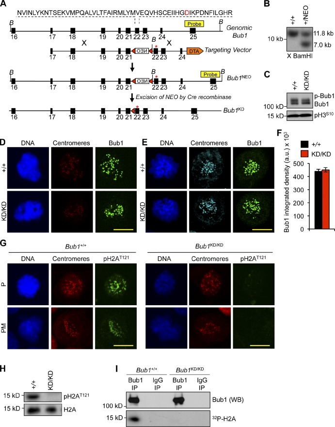
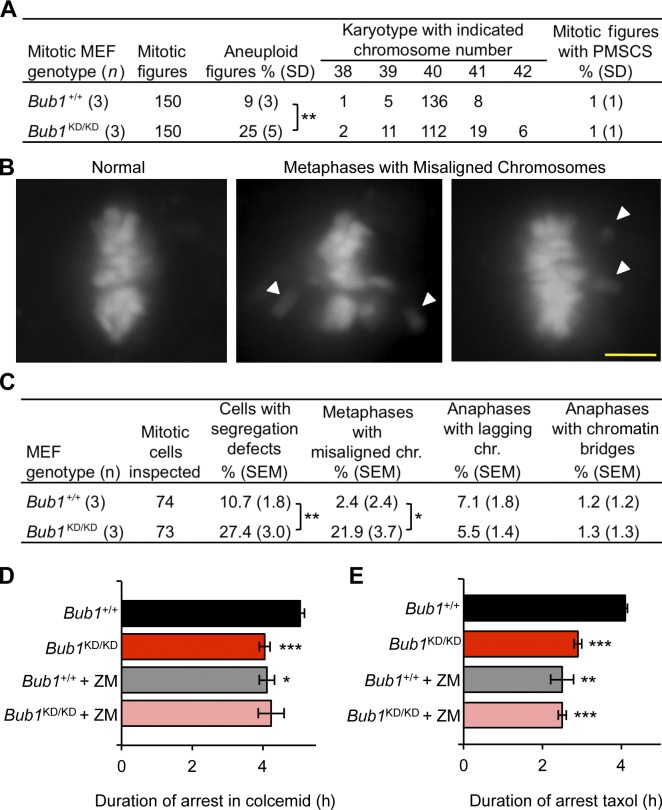
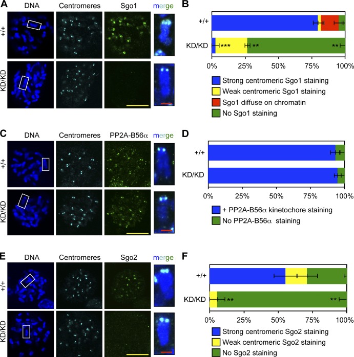
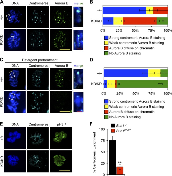
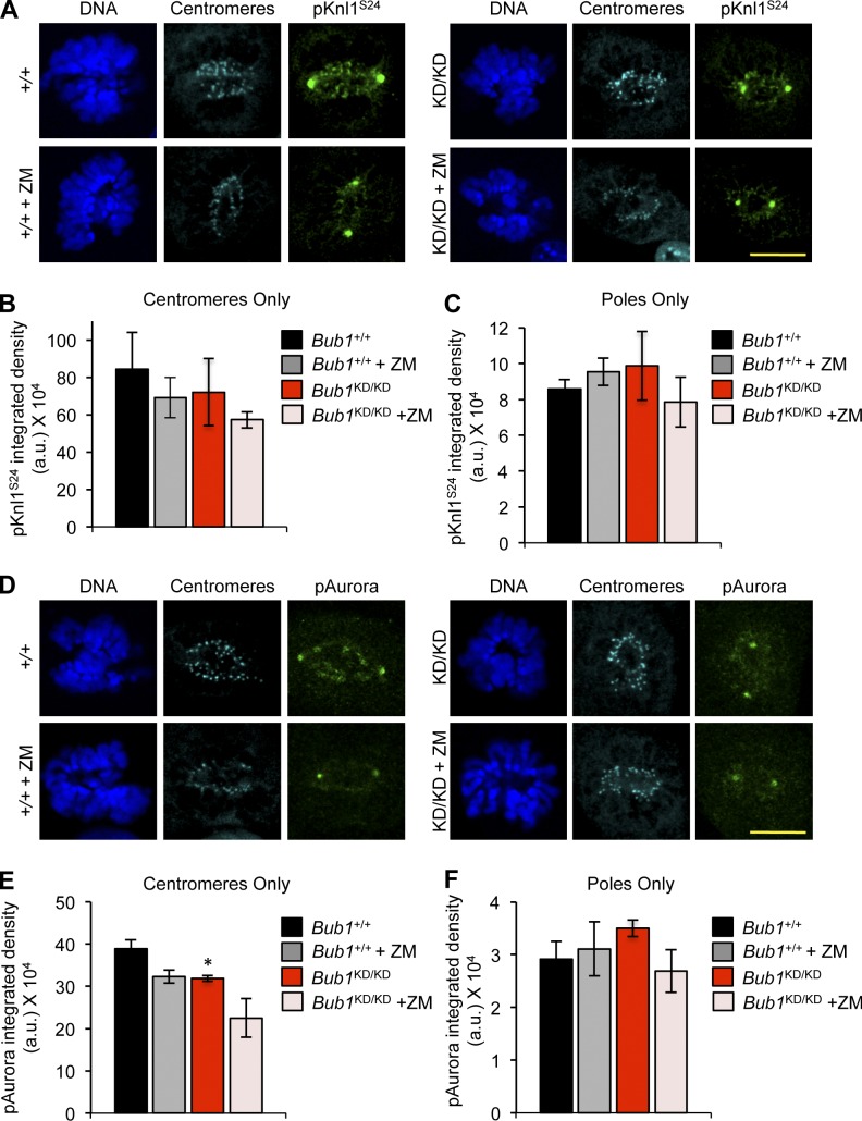
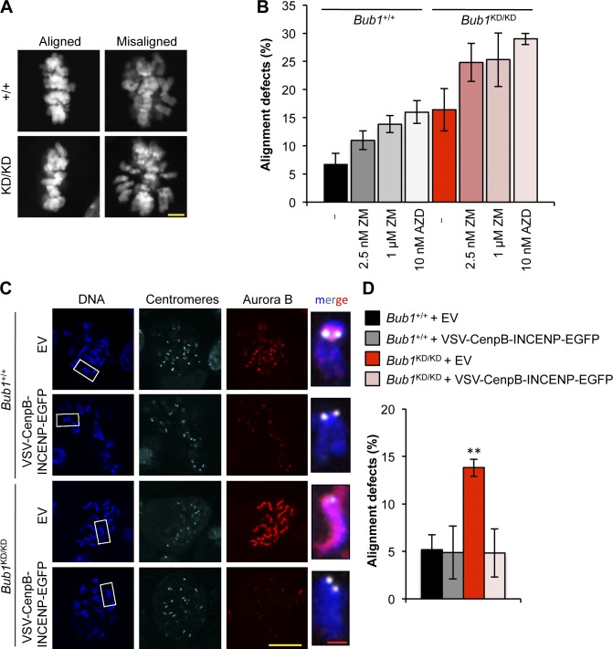
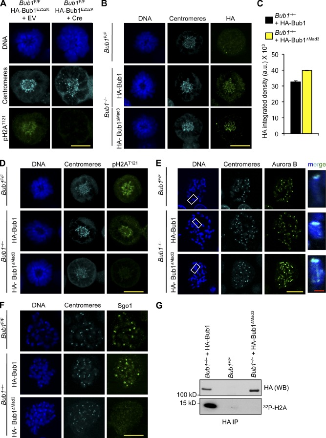
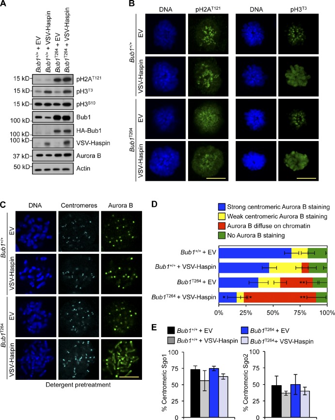
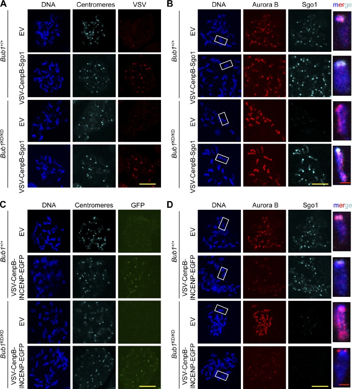
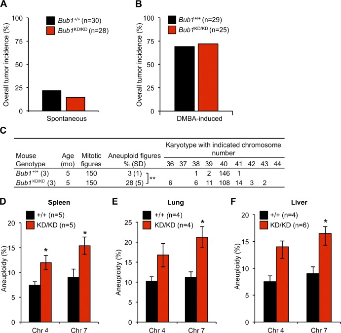
The mitotic checkpoint protein Bub1 is essential for embryogenesis and survival of proliferating cells, and bidirectional deviations from its normal level of expression cause chromosome missegregation, aneuploidy, and cancer predisposition in mice. To provide insight into the physiological significance of this critical mitotic regulator at a modular level, we generated Bub1 mutant mice that lack kinase activity using a knockin gene-targeting approach that preserves normal protein abundance. In this paper, we uncover that Bub1 kinase activity integrates attachment error correction and mitotic checkpoint signaling by controlling the localization and activity of Aurora B kinase through phosphorylation of histone H2A at threonine 121. Strikingly, despite substantial chromosome segregation errors and aneuploidization, mice deficient for Bub1 kinase activity do not exhibit increased susceptibility to spontaneous or carcinogen-induced tumorigenesis. These findings provide a unique example of a modular mitotic activity orchestrating two distinct networks that safeguard against whole chromosome instability and reveal the differential importance of distinct aneuploidy-causing Bub1 defects in tumor suppression.


Figures

Figure 1.
Figure 1.
Generation of Bub1 kinase–deficient mice. (A) Bub1 targeting strategy. Bub1 locus (+), D892N targeting vector, targeted allele (Bub1NEO), the targeted allele after Cre recombination (Bub1KD), BamHI (B) restriction sites, loxP sites (red triangles), and the Southern probe are indicated. DTA, diphtheria toxin A. Asterisks mark the D892N point mutation. (B) Southern blot of targeted ES clones digested with BamHI. X, digested. (C) Western blot of lysates from nocodazole-treated shake-off MEFs probed for Bub1 and pH3S10. (D) Immunostaining of Bub1+/+ and Bub1KD/KD MEFs. (E) Immunostaining of monastrol-treated Bub1+/+ and Bub1KD/KD MEFs. (F) Quantification of Bub1 staining shown in E from three independent MEF lines. Error bars indicate SEM. (G) Immunostaining of Bub1+/+ and Bub1KD/KD MEFs. (H) Western blot of lysates from taxol blocked MEFs probed for pH2AT121 and histone H2A. (I) Bub1 was immunoprecipitated from taxol-blocked Bub1+/+ and Bub1KD/KD immortalized MEFs and incubated with histone H2A in the presence of γ-[32P]ATP. a.u., arbitrary unit; NEO, neomycin; KD, kinase dead; IP, immunoprecipitation; WB, Western blot. Bars, 10 µm.
Figure 2.
Figure 2.
Bub1 kinase activity is required for chromosome alignment and maintenance of chromosomal stability. (A) Karyotype analysis of passage 5 MEFs. Statistics indicate comparison with wild type. (B) Representative images for chromosome segregation defects by live-cell imaging. White arrowheads shows misaligned chromosomes. Bar, 10 µm. (C) Live-cell imaging of chromosome segregation defects in primary MEFs. Statistics indicate comparison with wild type. (D) Analysis of mitotic duration in Bub1+/+ and Bub1KD/KD MEFs after colcemid addition (n = 3 or more lines per genotype). Error bars indicate SEM. The time at which 50% of the cells exited mitosis was compared. Statistics indicate comparison with wild type. (E) Same as D but with 0.5 µM taxol. chr., chromosome. *, P < 0.5; **, P < 0.1; ***, P < 0.001.
Figure 3.
Figure 3.
Optimal loading of shugoshins at inner centromeres requires Bub1 kinase activity. (A) IF of spreads from colcemid-blocked MEFs. Insets show enlarged chromosomes from boxed region (merged). (B) Quantification of Sgo1 localization (n = 3 lines per genotype). Error bars indicate SEM. Statistics indicate comparison with wild type. (C) Same as A except with PP2A-B56α. (D) Same as B except with PP2A-B56α. (E) Same as A except with Sgo2 (green). (F) Same as B except with Sgo2. **, P < 0.1; ***, P < 0.001. KD, kinase dead. Bars: (yellow) 10 µm; (red) 1 µm.
Figure 4.
Figure 4.
Bub1 kinase activity contributes to Aurora B accumulation at inner centromeres. (A) IF of spreads from unperturbed MEFs. Insets shows enlarged chromosomes from the boxed region (merged). Statistics indicate comparison with wild type. (B) Quantification of Aurora B localization (n = 3 MEF cell lines per genotype). Error bars indicate SEM. Statistics indicate comparison with wild type. (C) Same as A except with detergent pretreatment. (D) Same as B except with detergent pretreatment. (E) IF of mitotic MEFs. (F) Quantification of pH3T3 centromeric enrichment (n = 3 lines per genotype). Error bars indicate SEM. Statistics indicate comparison with wild type. *, P < 0.5; **, P < 0.1. KD, kinase dead. Bars: (yellow) 10 µm; (red) 1 µm.
Figure 5.
Figure 5.
Loss of Bub1 catalysis reduces inner centromeric Aurora B activity. (A) IF of mitotic MEFs after treatment with 1 µM ZM for 3 h. (B) Quantification of centromeric region (n = 3 MEF lines per genotype). Error bars indicate SEM. (C) Same as B except signal at spindle poles is quantified. (D) Same as A except with p-Aurora. Note that p-Aurora is a pan-Aurora antibody, and signal at poles is p-Aurora A, whereas signal at inner centromeres is p-Aurora B. (E) Same as B except with p-Aurora. Statistics indicate comparison with wild type without ZM. (F) Same as C but with p-Aurora. a.u., arbitrary unit; KD, kinase dead. *, P < 0.5. Bars, 10 µm.
Figure 6.
Figure 6.
Deregulated Aurora B activity promotes chromosome alignment defects in Bub1 kinase–deficient cells. (A) Representative images for cells with aligned or misaligned chromosomes after monastrol washout. (B) Analysis of chromosome alignment in cells that were treated with monastrol for 1 h and then with monastrol and MG132 for 1 h and released for 90 min into MG132 alone, with ZM or AZD (n = 3 independent MEF lines with ≥75 cells per group). Error bars indicate SEM. (C) IF of chromosome spreads from the indicated cells. Insets show enlarged chromosome from the boxed regions. (D) Analysis of chromosome alignment in cells released from monastrol as in B. Statistics indicate comparison with wild type with empty vector. EV, empty vector; KD, kinase dead. **, P < 0.1. Bars: (yellow) 10 µm; (red) 1 µm.
Figure 7.
Figure 7.
The N terminus of Bub1 is required for histone H2A phosphorylation. (A) IF of monastrol-blocked MEFs 4 d after infection with indicated viruses. (B) IF of monastrol-blocked MEFs. (C) Quantification of the HA signal, normalized from three independent experiments, is shown. Error bars indicate SEM. (D) IF of monastrol-blocked MEFs. (E) IF of spreads from colcemid-blocked MEFs. Staining was performed on spreads directly fixed in paraformaldehyde. Insets show enlarged chromosomes from the boxed regions. (F) IF of spreads from colcemid-blocked MEFs. (G) In vitro kinase assay for HA-Bub1 and HA-Bub1-ΔMad3. a.u., arbitrary unit; EV, empty vector; IP, immunoprecipitation; WB, Western blot. Bars: (yellow) 10 µm; (red) 1 µm.
Figure 8.
Figure 8.
Phosphorylated histone H3T3 and histone H2AT121 together serve as the minimal binding element for stable Aurora B association. (A) Western blot of lysates from MEF extracts. Actin was used as a loading control. (B) IF of monastrol-blocked MEFs. (C) IF of spreads from colcemid-blocked MEFs. Staining was performed on spreads that were first treated with detergent before fixation. (D) Quantification of Aurora B (n = 3 lines per group). Staining was performed on spreads with detergent before fixation. Statistics indicate comparison with wild type with empty vector. (E) Quantification of cells with centromeric Sgo1 or Sgo2. Error bars indicate SEM. EV, empty vector. *, P < 0.5; **, P < 0.1. Bars, 10 µm.
Figure 9.
Figure 9.
Inner centomeric recruitment of Aurora B and Sgo1 lacks interdependence in the absence of Bub1 kinase activity. (A) IF of spreads from colcemid-blocked MEFs. (B) IF of spreads from colcemid-blocked MEFs. Chromosomes were stained for Aurora B (red), Sgo1 (cyan), and DNA (blue). Staining was performed on spreads directly fixed in paraformaldehyde. Insets shows enlarged chromosomes from boxed regions (merged). (C) IF of spreads from colcemid blocked MEFs. (D) IF of spreads from colcemid blocked MEFs. Staining was performed on spreads directly fixed in paraformaldehyde. Insets show enlarged chromosomes from boxed regions (merged). EV, empty vector. Bars: (yellow) 10 µm; (red) 1 µm.
Figure 10.
Figure 10.
Bub1 kinase activity is required for protection from aneuploidization but not tumor suppression. (A) Spontaneous tumor incidence of 14-mo-old mice. (B) Overall tumor incidence of DMBA-treated mice. (C) Karyotype analysis of splenocytes from 5-mo-old mice. (D–F) Quantification of chromosome (Chr) 4 and 7 copies by interphase FISH. Statistics indicate comparison with wild type. KD, kinase dead. Error bars indicate SEM. *, P < 0.5; **, P < 0.1.

Similar articles

Cited by

References

    1. Babu J.R., Jeganathan K.B., Baker D.J., Wu X., Kang-Decker N., van Deursen J.M. 2003. Rae1 is an essential mitotic checkpoint regulator that cooperates with Bub3 to prevent chromosome missegregation. J. Cell Biol. 160:341–353 10.1083/jcb.200211048 - DOI - PMC - PubMed
    1. Baker D.J., Jeganathan K.B., Cameron J.D., Thompson M., Juneja S., Kopecka A., Kumar R., Jenkins R.B., de Groen P.C., Roche P., van Deursen J.M. 2004. BubR1 insufficiency causes early onset of aging-associated phenotypes and infertility in mice. Nat. Genet. 36:744–749 10.1038/ng1382 - DOI - PubMed
    1. Baker D.J., Jeganathan K.B., Malureanu L., Perez-Terzic C., Terzic A., van Deursen J.M. 2006. Early aging–associated phenotypes in Bub3/Rae1 haploinsufficient mice. J. Cell Biol. 172:529–540 10.1083/jcb.200507081 - DOI - PMC - PubMed
    1. Baker D.J., Jin F., Jeganathan K.B., van Deursen J.M. 2009. Whole chromosome instability caused by Bub1 insufficiency drives tumorigenesis through tumor suppressor gene loss of heterozygosity. Cancer Cell. 16:475–486 10.1016/j.ccr.2009.10.023 - DOI - PMC - PubMed
    1. Bolanos-Garcia V.M., Kiyomitsu T., D’Arcy S., Chirgadze D.Y., Grossmann J.G., Matak-Vinkovic D., Venkitaraman A.R., Yanagida M., Robinson C.V., Blundell T.L. 2009. The crystal structure of the N-terminal region of BUB1 provides insight into the mechanism of BUB1 recruitment to kinetochores. Structure. 17:105–116 10.1016/j.str.2008.10.015 - DOI - PMC - PubMed

Publication types

MeSH terms