. 2012 Oct 31;45(5):241-50.
doi: 10.1267/ahc.12013. Epub 2012 Sep 22.

Anatomy and histology of rodent and human major salivary glands: -overview of the Japan salivary gland society-sponsored workshop-

Affiliations

Anatomy and histology of rodent and human major salivary glands: -overview of the Japan salivary gland society-sponsored workshop-

Osamu Amano et al. Acta Histochem Cytochem. .

Abstract

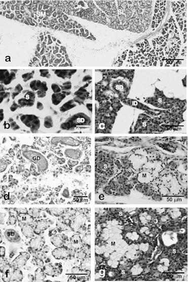
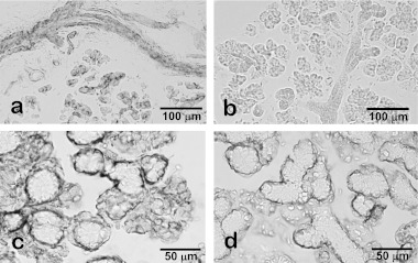
MAJOR SALIVARY GLANDS OF BOTH HUMANS AND RODENTS CONSIST OF THREE PAIRS OF MACROSCOPIC GLANDS: parotid, submandibular, and sublingual. These glands secrete serous, mucous or mixed saliva via the proper main excretory ducts connecting the glandular bodies with the oral cavity. A series of discoveries about the salivary ducts in the 17th century by Niels Stensen (1638-1686), Thomas Wharton (1614-1673), and Caspar Bartholin (1655-1738) established the concept of exocrine secretion as well as salivary glands. Recent investigations have revealed the endocrine functions of parotin and a variety of cell growth factors produced by salivary glands.The present review aims to describe macroscopic findings on the major salivary glands of rodents and the microscopic differences between those of humans and rodents, which review should be of interest to those researchers studying salivary glands.

Keywords: human; immunohistochemistry; mouse; rat; salivary glands.


Figures

Fig. 1
Fig. 1
Macroscopic anatomy of the anterior and lateral neck portions of mice before (a) and after (b, c) the perfusion of fixative and removal of fat tissues. Anterior neck is occupied by large white fat tissues including salivary glands (a). Upon removing fat tissues and submandibular lymph nodes, salivary and extraorbital lacrimal glands were exposed. The extraorbital lacrimal gland (*) is located anterior to the parotid gland (P). The submandibular (SM) and sublingual (SL) glands are encapsulated with common fascia.
Fig. 2
Fig. 2
Electron microscopy of striated duct cells of the submandibular gland (a) and distal urinary tubule cells of the kidney (b) of rats. Both cells develop numerous vertically-arranged mitochondria (*) and the basal infoldings of cell membrane (the basal striation).
Fig. 3
Fig. 3
Immunohistochemistry and electron microscopy of the intercalated duct of the rat submandibular gland. Smooth muscle-action-immunohistochemistry shows distribution of myoepithelial cells along the intercalated duct (*) as well as acini (A). No myoepithelial cells are associated with other duct portions such as the striated (SD) and granular ducts (GD). The intercalated duct cells are cuboidal in shape and partially surrounded by myoepithelial cells (arrowhead, b). Hsp27-immunohistochemistry of 3-week-old rat submandibular gland shows the centrally-located immunopositive terminal tubule cells (arrows, d). Electron micrograms of Hsp27-immunohistochemistry of 4-week-old rat submandibular gland show that Hsp27-immunoreactive terminal tubule cells differentiated into immature acinar cells (d) and granular intercalated duct cells (e). L, lumen; S, secretory granules in acinar cells.
Fig. 4
Fig. 4
Photomicrographs of rat (a, b, e, f), mouse (c, d) and human (g) submandibular glands. Electron microscopy shows supranuclear localization of numerous exocrine secretory granules in the granular duct cells (a). FGF2-immunoelectron microscopy shows an immunopositive pillar cell (*) without large secretory granules present between the principal granular duct cells without FGF2-immunoreactivity (G) (b). EGF-immunohistochemistry of the submandibular glands shows specific localization of EGF in the granular duct in male (c) and female (d) mouse. Note that EGF-immunopositive granular ducts are less developed in the female submandibular glands than the male glands. Colloidal-gold particles representing the immunoreactivity for EGF are exclusively localized in the secretory granules of the granular duct cells (e). Expression of EGF-mRNA is specifically localized in the basal cytoplasm of the granular duct cells (f). In human submandibular glands, EGF-immunoreactivity is dominantly localized in the striated duct (arrows) and partially in the serous acinar cells (arrowheads). No immunoreaction is detected in mucous acini (*) (g).
Fig. 5
Fig. 5
Light micrographs of major salivary glands of the rat (a, b, d, f) and human (c, e, g). Major salivary glands of rats are easily distinguishable light-microscopically by the histological features of acinar and ductal structures (a). In rat (b) and human (c) parotid glands, acini are composed of serous (or seromucous) cells. In rat submandibular glands (d), the granular ducts (GCT) are markedly developed but mucous acini and serous demilunes are not recognized. In human submandibular glands (e), mixed acini accompanying serous demilunes are observed whereas no GCT portions are found. In rat (f) and human (g) sublingual glands, mixed acini accompanying the serous demilunes are observed. P, parotid gland; SL, sublingual gland; SM, submandibular gland; S, serous acini; M, mucous acini; SD, striated duct; ID, intercalated duct; GD, granular duct; D, demilune.
Fig. 6
Fig. 6
Non-specific immunoreactions in the rodent salivary glands. Without specific primary antibodies anti-mouse IgG secondary antibody reacted with duct cells and luminal membrane of acinar cells in rat parotid glands (a) whereas no non-specific reaction was detected by using pre-absorbed secondary antibody for rat tissues (b). The immunohistochemical procedure for the rat sublingual glands by the combination of an anti-smooth muscle actin mouse monoclonal antibody, an established marker for myoepithelial cells, and conventional anti-mouse IgG antibody triggers a broader immunoreaction than expected, including serous demilunes (c). After replacing the secondary antibody with the pre-absorbed one for rat tissue, the immunoreaction is confirmed to localize in myoepithelial cells (d).

References

    1. Aloe L., Alleva E., Böhm A., Levi-Montalcini R. Aggressive behavior induces release of nerve growth factor from mouse salivary gland into the bloodstream. Proc. Natl. Acad. Sci. U S A. 1986;83:6184–6187. - PMC - PubMed
    1. Amano O. The salivary gland: anatomy for surgeons and researchers. Jpn. J. Oral Maxillofac. Surg. 2011;57:384–393.
    1. Amano O., Tsuji T., Nakamura T., Iseki S. Expression of transforming growth factor beta-1 in the submandibular gland of the rat. J. Histochem. Cytochem. 1991;39:1707–1711. - PubMed
    1. Amano O., Iseki S. Expression, localization and developmental regulation of insulin-like growth factor I mRNA in rat submandibular gland. Archs. Oral Biol. 1993;38:671–677. - PubMed
    1. Amano O., Yoshitake Y., Nishikawa K., Iseki S. Basic fibroblast growth factor in rat salivary glands. Cell Tissue Res. 1993;273:467–474. - PubMed