. 2012;10(11):e1001432.
doi: 10.1371/journal.pbio.1001432. Epub 2012 Nov 27.

Structural mechanism of ER retrieval of MHC class I by cowpox

Affiliations

Structural mechanism of ER retrieval of MHC class I by cowpox

William H McCoy 4th et al. PLoS Biol. 2012.

Abstract

One of the hallmarks of viral immune evasion is the capacity to disrupt major histocompatibility complex class I (MHCI) antigen presentation to evade T-cell detection. Cowpox virus encoded protein CPXV203 blocks MHCI surface expression by exploiting the KDEL-receptor recycling pathway, and here we show that CPXV203 directly binds a wide array of fully assembled MHCI proteins, both classical and non-classical. Further, the stability of CPXV203/MHCI complexes is highly pH dependent, with dramatically increased affinities at the lower pH of the Golgi relative to the endoplasmic reticulum (ER). Crystallographic studies reveal that CPXV203 adopts a beta-sandwich fold similar to poxvirus chemokine binding proteins, and binds the same highly conserved MHCI determinants located under the peptide-binding platform that tapasin, CD8, and natural killer (NK)-receptors engage. Mutagenesis of the CPXV203/MHCI interface identified the importance of two CPXV203 His residues that confer low pH stabilization of the complex and are critical to ER retrieval of MHCI. These studies clarify mechanistically how CPXV203 coordinates with other cowpox proteins to thwart antigen presentation.


Conflict of interest statement

The authors have declared that no competing interests exist.

Figures

Figure 1
Figure 1. CPXV203 downregulation of MHCI is β2m-dependent but PLC-independent.
(A) Hemagglutinin-tagged CPXV203 (CPXV203-HA) stably expressed in β2m−/− cells was unable to co-IP HC. (B) MEFs lacking TAP/Tpn were transduced with CPXV203/CPXV012-IRES-GFP and then MHCI surface expression was monitored by flow-cytometry. B6-derived MEF cell lines expressing TAP/Tpn typically have a mean fluorescence of MHCI surface expression of around 200 as shown in the CRT add-back control in (C), lower left. Data in (B) demonstrate that CPXV203 does not require PLC components to downregulate MHCI, while CPXV012 is TAP/Tpn-dependent. (C) Extension of the studies in B to CRT−/− cells showed that CPXV203 function is independent of CRT. (D) Stable expression of CPXV203 did not impair TAP/Tpn association as monitored by co-IP, and CPXV203 did not reduce steady-state levels of CRT found in cell lysate. Numbers at the top of dot plots in (B) and (C) indicate the mean fluorescence intensity of GFP-negative and -positive populations.
Figure 2
Figure 2. CPXV203 binds MHCI with higher affinity at low pH.
(A,B) SPR analysis of CPXV203/Kb binding at pHER 7.4 (A) and pHGolgi 6.0 (B). Neutravidin was used to capture site-specifically biotinylated MHCI prior to injection of increasing concentrations of CPXV203. Non-specific binding was addressed via reference subtraction of a neutravidin-only control flow cell. SPR curves (grey) were fit kinetically using a 1∶1 Langmuir model (black lines). See Table S1 for additional MHCI alleles and Figure S1 for analysis of protein oligomeric state. CPXV203 did not bind β2m alone (pH 6.0/7.4, unpublished data). (C) Equilibrium BLI analysis confirmed CPXV203 affinity increased for murine/primate MHC Ia/Ib alleles as pH decreased from 7.6 to 6.0. Neutravidin sensors captured site-specifically biotinylated MHCI prior to immersion in samples of varying pH with constant [CPXV203]. The [CPXV203] chosen for this experiment produced the lowest detectable signal at pHER 7.4 for each MHCI. Nonspecific binding was addressed using both reference subtraction (neutravidin) and standard blocking reagents (1% BSA +0.05% TWEEN). *Indicates complex includes murine β2m instead of human β2m.
Figure 3
Figure 3. Crystal structure of CPXV203 bound to MHCI.
(A) Ribbon diagram of the complex structure of CPXV203 (magenta), Kb (blue), β2m (grey), and OVA257–264 (yellow, spheres). No N-linked glycosylation sites are present near the interface. Membrane proximal domain shifts, α3 (7.2°) and β2m (16.8°), are within the range observed in previous crystal structures of free MHCI (see Text S1). (B) Comparison of CPXV203/MHCI binding orientation to other MHCI binders: viral (US2) , chaperone (Tpn), co-stimulatory (CD8αα) , and NK receptor (Ly49C) . Chains colored as in A. Proposed Tpn contact loops (α2 128–136, α3 222–229) are colored magenta with contacts identified by mutagenesis shown as spheres ,,,. See also Table S2 and Figure S2.
Figure 4
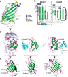
Figure 4. Structural topology of CPXV203 and comparison to poxvirus CKBPs.
(A–D) CPXV203 regions used to contact α2, α3, and β2m domains are indicated. (A) Ribbon diagram of CPXV203 (R5-S190) colored according to 2° structure (cyan α helices, blue 310 helices, green β strands, grey loops, and yellow disulfide bonds). CPXV203 orientation is identical to Figure 3A. (B) Topology diagram of CPXV203 with 2° structure coloring as in (A). Disulfide bonds are shown as flattened balls-and-sticks with residue positions listed. The core β sandwich is divided into its β sheets by a dashed, grey line. Structural elements not found in vCCI (PDB: 1CQ3) are highlighted in black, including the absence of a highly negatively charged chemokine-binding (CKB) loop. MHCI α2 and β2m contacts are highly localized to these unique structural elements. (C) CPXV203 and poxvirus CKBPs (RPV vCCI and ECTV CrmD-CTD) use three distinct surfaces for ligand binding. Each CKBP/chemokine (CK) complex (PDBs: 3ONA and 2FFK [54]) was aligned to CPXV203 using CE . The view from Figure 3A has been rotated −90° (y-axis). Complexes are shown as ribbon diagrams: CPXV203 and CKBP (magenta), core β-sandwich (green), H-2Kb (blue), β2m (grey), OVA257–264 (yellow, spheres), CK (cyan). (D) CPXV203 and CKBPs are shown after a further −90° y-axis rotation to highlight the distinct β7–β9 junction found in CPXV203 and CrmD-CTD relative to vCCI-like proteins. The absence of a CKB loop in CPXV203 and CrmD-CTD relative to vCCI is also indicated. See also Tables S3, S4, and Figure S3.
Figure 5
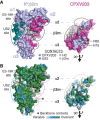
Figure 5. CPXV203 binds conserved elements within each MHCI domain.
The CPXV203/MHCI complex is shown as a Connolly surface (1.4 Å probe). Relative to Figure 3A, MHCI was rotated −30° (y-axis), while CPXV203 was rotated 60° (y-axis). Each domain-specific interaction is circled on both molecules. The US2 and E3-19K binding sites are also indicated. (A) CPXV203/MHCI surfaces are colored by chain (see Figure 3A) or by contact (see legend). CPXV203/MHCI interfaces are labeled according to the contacted MHCI domain. Each MHCI domain is contacted by a CPXV203 surface that is localized to a distinct structural region. Comparison of the CPXV203 and US2/E3-19K sites clearly shows the lack of binding site overlap between PLC-proximal and PLC-distal immune evasion proteins. (B) Conservation of interface residues is shown for CPXV203-susceptible MHCI and T4 poxvirus proteins (see Figures S4A and S3A, respectively). Backbone contacts for each interface are indicated (*). CPXV203 contacts highly conserved MHCI surfaces, as opposed to US2 (prominent variable residues between α2–α3). Conservation of MHCI contact residues in the T4 family are highly localized to the α3 interface, which contains 4/6 CPXV203 backbone contact positions. See also Figure S4 and Table S4 and S5.
Figure 6
Figure 6. Mutagenesis supports a tri-domain interface and pH regulation.
(A–C) 3KO cells stably transduced with β2m and Kb (3KO-β2m-Kb) were transduced with CPXV203-IRES-GFP to evaluate the effect of CPXV203/MHCI interface mutation on surface Kb remaining relative to untransduced cells. Comparable expression of wild-type and mutant proteins was determined using co-expressed GFP and/or western blotting. Each figure represents the data from two independent experiments. **p<0.01; ***p<0.001 when compared with wild type. Limited MHCI escape was observed for two single α3 interface mutations (Kb E229Y or CPXV203 F76A), while significant Kb escape required double mutation of the α3 interface (Kb D227K/E229Y, CPXV203 H75A/H80A, Kb Q226A/CPXV203 F76A, Kb E229Y/CPXV203 H75A, Kb E229Y/CPXV203 F76A) or simultaneous mutation of interfaces α2 and α3 (CPXV203 F76A, Y161A) (D) Most mutant CPXV203-HA exhibited a decreased ability to co-IP Kb even though surface Kb (%) did not increase significantly. CPXV203/Kb mutations that significantly increased surface Kb expression also exhibited a decrease in total Kb (lysate), indicative of the release of MHCI retention . (E) Kinetic analysis (SPR) of alanine mutation of α3 interface histidines (CPXV203 H75 and H80) summarized by on-rate (ka, (+)-axis) and half-life (half-life, (−)-axis). (F) Summary of mutagenic analysis (functional and biosensor) mapped to CPXV203/MHCI structure (same colors as Figure 3A) shown as cartoon loops except for the peptide (spheres). Mutated residues (Cα spheres) were colored to indicate whether they had a significant effect (red) or no significant effect (grey). CPXV203 and MHCI have been translated apart (x-axis) to highlight corresponding interface mutations. See also Tables S6, S7.
Figure 7
Figure 7. CPXV203 co-opts the KDELR to retrieve MHCI from the Golgi.
Comparison of the separate but complementary strategies employed by CPXV012 and CPXV203 to downregulate MHCI. CPXV012 impairs nascent MHCI folding by inhibiting TAP, while CPXV203 selectively binds mature MHCI in the low pH environment found in the ERGIC-Golgi and then uses the KDELR to return to the ER where KDELR/CPXV203/MHCI can rapidly dissociate.

Comment in

  • How cowpox virus turns back cell defenses.
    Sedwick C. Sedwick C. PLoS Biol. 2012;10(11):e1001431. doi: 10.1371/journal.pbio.1001431. Epub 2012 Nov 27. PLoS Biol. 2012. PMID: 23209376 Free PMC article. No abstract available.

Similar articles

Cited by

References

    1. Hansen TH, Bouvier M (2009) MHC class I antigen presentation: learning from viral evasion strategies. Nat Rev Immunol 9: 503–513. - PubMed
    1. Lilley BN, Ploegh HL (2005) Viral modulation of antigen presentation: manipulation of cellular targets in the ER and beyond. Immunol Rev 207: 126–144. - PubMed
    1. Früh K, Ahn K, Djaballah H, Sempé P, van Endert PM, et al. (1995) A viral inhibitor of peptide transporters for antigen presentation. Nature 375: 415–418. - PubMed
    1. Hill A, Jugovic P, York I, Russ G, Bennink J, et al. (1995) Herpes simplex virus turns off the TAP to evade host immunity. Nature 375: 411–415. - PubMed
    1. Ahn K, Gruhler A, Galocha B, Jones TR, Wiertz EJ, et al. (1997) The ER-luminal domain of the HCMV glycoprotein US6 inhibits peptide translocation by TAP. Immunity 6: 613–621. - PubMed

Publication types

MeSH terms