. 2012;8(11):e1003047.
doi: 10.1371/journal.ppat.1003047. Epub 2012 Nov 29.

Neutrophil-derived IL-1β is sufficient for abscess formation in immunity against Staphylococcus aureus in mice

Affiliations

Neutrophil-derived IL-1β is sufficient for abscess formation in immunity against Staphylococcus aureus in mice

John S Cho et al. PLoS Pathog. 2012.

Abstract

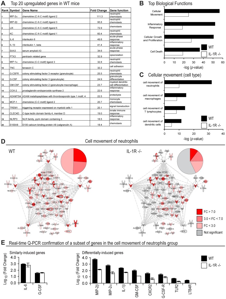
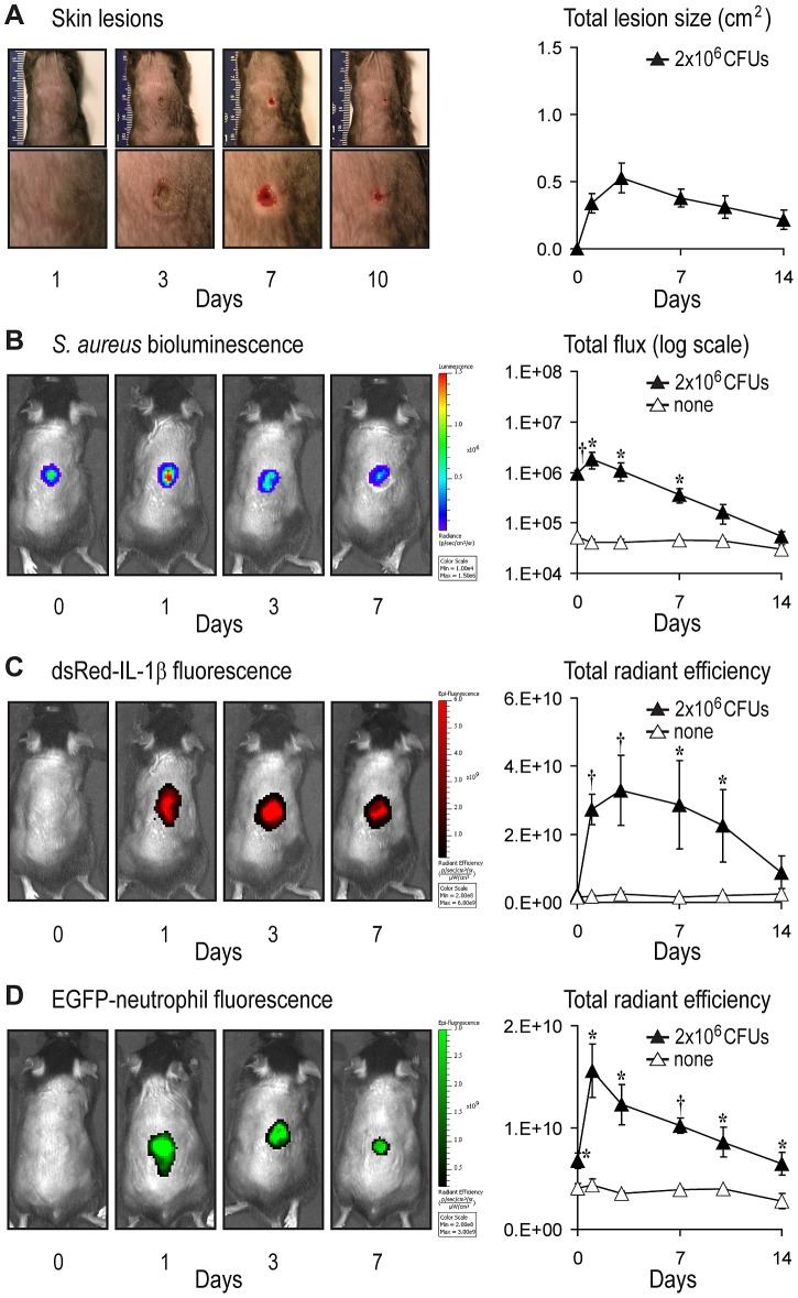
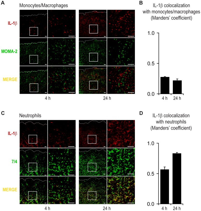
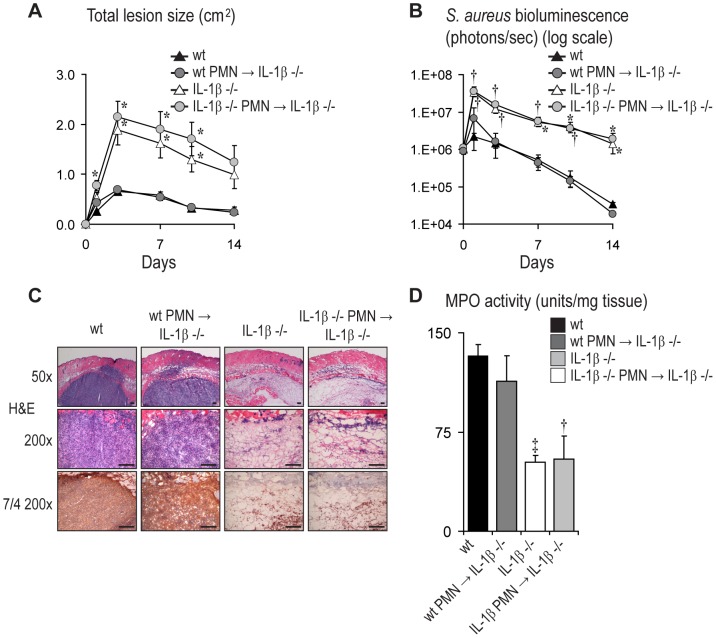
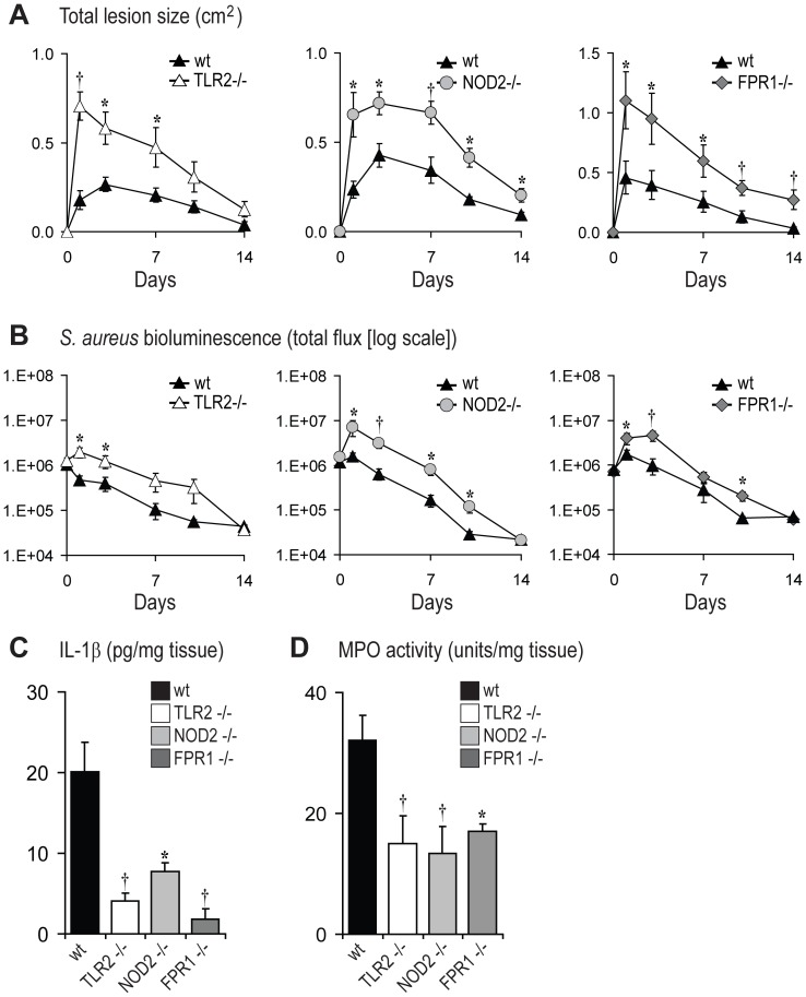
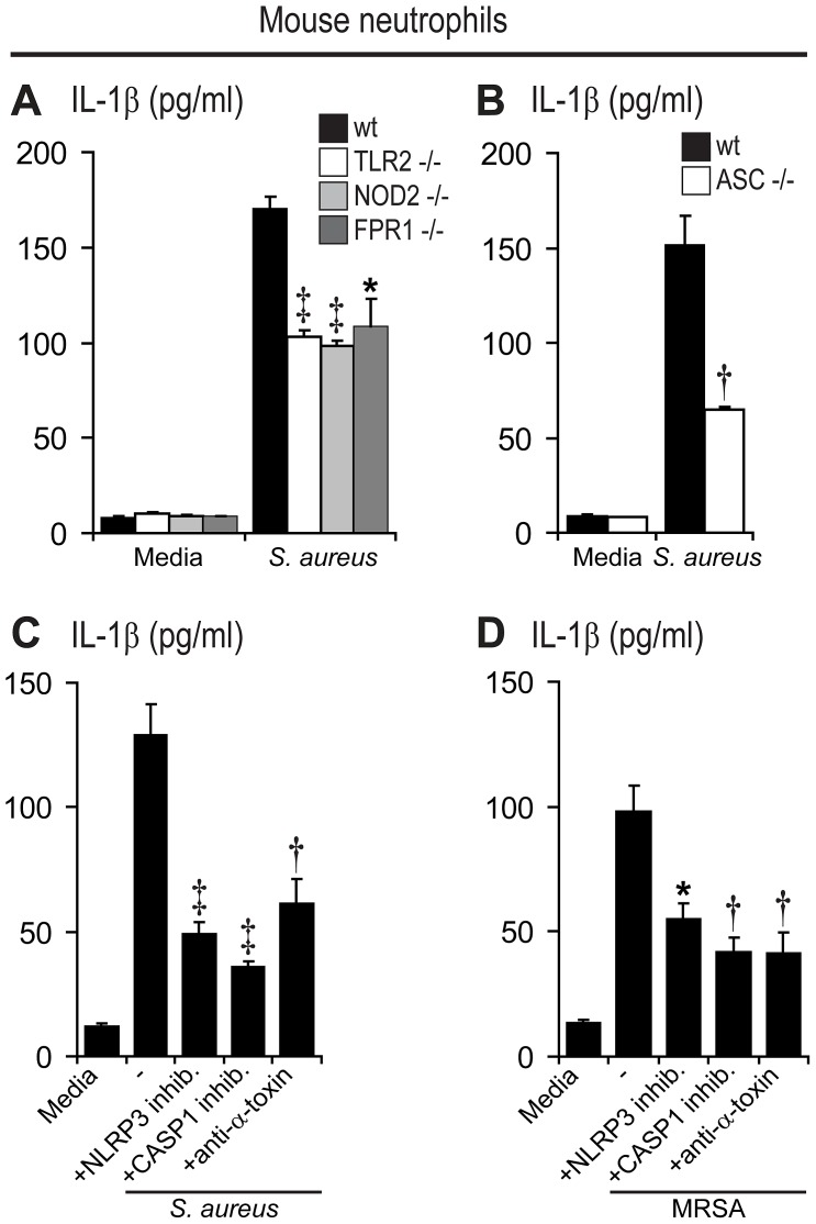
Neutrophil abscess formation is critical in innate immunity against many pathogens. Here, the mechanism of neutrophil abscess formation was investigated using a mouse model of Staphylococcus aureus cutaneous infection. Gene expression analysis and in vivo multispectral noninvasive imaging during the S. aureus infection revealed a strong functional and temporal association between neutrophil recruitment and IL-1β/IL-1R activation. Unexpectedly, neutrophils but not monocytes/macrophages or other MHCII-expressing antigen presenting cells were the predominant source of IL-1β at the site of infection. Furthermore, neutrophil-derived IL-1β was essential for host defense since adoptive transfer of IL-1β-expressing neutrophils was sufficient to restore the impaired neutrophil abscess formation in S. aureus-infected IL-1β-deficient mice. S. aureus-induced IL-1β production by neutrophils required TLR2, NOD2, FPR1 and the ASC/NLRP3 inflammasome in an α-toxin-dependent mechanism. Taken together, IL-1β and neutrophil abscess formation during an infection are functionally, temporally and spatially linked as a consequence of direct IL-1β production by neutrophils.


Conflict of interest statement

The authors have declared that no competing interests exist.

Figures

Figure 1
Figure 1. Gene expression analysis in wt and IL-1R-deficient mice after S. aureus skin infection.
Wt and IL-1R−/− mice were inoculated intradermally with S. aureus and gene expression analysis was performed on skin specimens taken at 4 hrs and compared with samples from uninfected skin (n = 3 mice per group). Upregulated genes were defined according to the criteria of fold-change (FC) >1.5, p-value<0.05. (A) Top 20 induced genes in wt mice ordered by FC (gene function was assigned using NetAffx Gene Ontology Mining Tool [Affymetrix]). (B) Top four biological functions in wt versus IL-1R−/− mice generated by Ingenuity Pathway analysis (ranked by p-value). (C) Top four cell types in the Cellular Movement functional group (ranked by p-value). (D) Functional network analysis of genes in the Cell Movement of Neutrophils group in wt mice (left) compared with the same genes in IL-1R−/− mice (right). The genes in the networks and pie charts are shaded according to FC. Red: >7.0 FC (strongly upregulated), pink: 3.0<FC<7.0 (moderately upregulated), light pink: <3.0 FC (slightly upregulated), or grey: no change or not statistically significant. (E) Real-time Q-PCR confirmation (mean log10 fold change ± SEM) of 2 representative genes that were similarly-induced between wt and IL-1R-deficient mice and 8 representative genes that were differentially-induced in wt mice compared with IL-1R-deficient mice from the Cell Movement of Neutrophils sub-group from microarray analysis in Fig. 1D.
Figure 2
Figure 2. Real-time kinetics of IL-1β production and neutrophil recruitment after S. aureus skin infection.
pIL1-DsRed reporter mice or LysEGFP mice were inoculated intradermally with S. aureus. (A) Representative skin lesions (left) (entire dorsal backs [top, millimeter ruler shown for scale] and close-ups of lesions [bottom]) and mean total lesion size (cm2) ± SEM (right). (B) Representative in vivo S. aureus bioluminescence (left) and mean total flux (photons/s) ± SEM (logarithmic scale) (right). (C) Representative in vivo EGFP-neutrophil fluorescence (left) and mean total radiant efficiency (photons/s)/(µW/cm2) ± SEM (right). (D) Representative in vivo DsRed-IL-1β fluorescence (left) and mean total radiant efficiency (photons/s)/(µW/cm2) ± SEM (right). Data are from 2 experiments with at least 4 mice/group per experiment. *p<0.05; p<0.01, S. aureus-infected mice versus none (sham injection alone) (Student's t-test).
Figure 3
Figure 3. Neutrophils are the predominant cell type that expresses IL-1β after S. aureus skin infection.
pIL1-DsRed mice were infected intradermally with S. aureus and lesional skin specimens were collected at 4 and 24 hrs. Representative photomicrographs of sections labeled with anti-DsRed (IL-1β, red) and anti-MOMA2 (monocytes/macrophages, green) (A) or anti-7/4 (neutrophils, green) (C) and sections analyzed by confocal microscopy. Cells expressing both markers appear yellow (merge). High (left) and low (right) magnification images are shown (Scale bars = 50 µm). Dotted line = dermoepidermal junction. Quantification of co-localization of IL-1β-DsRed fluorescence with MOMA2+ monocytes/macrophages (B) or 7/4+ neutrophils (D) using the Manders' coefficient for a value range of 0 to 1 in which 0 = no pixels co-localize and 1 = all pixels co-localize. Data are representative from 4 mice per group.
Figure 4
Figure 4. Neutrophil-derived IL-1β is sufficient for abscess formation and bacterial clearance of S. aureus-infected mice.
Neutrophils from IL-1β−/− or wt donor mice were adoptively transferred into IL-1β−/− recipient mice. After 2 hrs, these mice and normal wt and IL-1β−/− mice were infected intradermally with S. aureus. (A) Mean total lesion size (cm2) ± SEM. (B) In vivo bioluminescence quantified by mean total flux (photons/s) ± SEM (logarithmic scale). (C) Representative photomicrographs of sections labeled with H&E stain and anti-7/4 (neutrophils) (immunoperoxidase method) at 1 day after inoculation. Scale bars = 100 µm. (D) Mean myeloperoxidase (MPO) activity (U/mg tissue weight) ± SEM from lesional skin at 1 day after inoculation. Data are representative from 2 independent experiments with at least 4 mice/group. *p<0.05; p<0.01, p<0.001, IL-1β−/− mice or mice with adoptively transferred neutrophils versus wt mice (Student's t-test).
Figure 5
Figure 5. The PRRs TLR2, NOD2, and FPR1 contribute to optimal production of IL-1β during infection.
TLR2−/−, NOD2−/−, FPR1−/− and wt mice were inoculated intradermally with S. aureus. (A) Mean total lesion size (cm2) ± SEM. (B) In vivo bioluminescence quantified by mean total flux (photons/s) ± SEM (logarithmic scale). (C) Mean IL-1β protein levels (pg/mg tissue weight) ± SEM from lesional skin at 4 hrs after inoculation. (D) Mean myeloperoxidase (MPO) activity (U/mg tissue weight) ± SEM from lesional skin at 4 hrs after inoculation. *p<0.05, p<0.01, TLR2−/−, NOD2−/− or FPR1−/− mice versus wt mice (Student's t-test). Data are from 2 experiments with at least 4 mice/group per experiment.
Figure 6
Figure 6. PRRs, α-toxin and the ASC/NLRP3 inflammasome contribute to S. aureus- and MRSA-induced IL-1β production by mouse neutrophils.
Neutrophils from mouse bone marrow were infected with live S. aureus (SH1000) or MRSA (USA300 LAC strain) (MOI bacteria∶neutrophils of 5∶1) for a total culture time of 6 hrs and gentamicin was added at 60 min from the start of the infection to prevent bacterial overgrowth. IL-1β protein levels (mean ± SEM) were measured in culture supernatants by ELISA. (A, B) IL-1β protein levels in supernatants from S. aureus-infected neutrophils from TLR2−/−, NOD2−/−, FPR1−/−, ASC−/− and wt mice. (C, D) IL-1β protein levels in supernatants from wt mouse neutrophils infected with S. aureus (C) or MRSA (D), in the absence and presence of an NLRP3-inhibitor (glibenclamide), a caspase-1 inhibitor (Z-YVAD-FMK) or anti-staphylococcal α-toxin antiserum. The respective vehicle controls (DMSO or normal rabbit IgG) for the inhibitors had IL-1β protein levels that did not differ than media alone (data not shown). Data are from 3–5 mice per group. *p<0.05; p<0.01, p<0.001, TLR2−/−, NOD2−/−, FPR1−/− or ASC−/− mice versus wt mice (A, B) or the NLRP3-inhibitor, caspase-1 inhibitor or anti-staphylococcal α-toxin antiserum treatment versus media alone (C, D) (Student's t-test).

References

    1. Borregaard N (2010) Neutrophils, from marrow to microbes. Immunity 33: 657–670 S1074-7613(10)00417-6 [pii];10.1016/j.immuni.2010.11.011 [doi]. - PubMed
    1. Rigby KM, Deleo FR (2012) Neutrophils in innate host defense against Staphylococcus aureus infections. Semin Immunopathol 34: 237–259 10.1007/s00281-011-0295-3 [doi]. - PMC - PubMed
    1. Miller LS, O'Connell RM, Gutierrez MA, Pietras EM, Shahangian A, et al. (2006) MyD88 mediates neutrophil recruitment initiated by IL-1R but not TLR2 activation in immunity against Staphylococcus aureus . Immunity 24: 79–91. - PubMed
    1. Daum RS (2007) Clinical practice. Skin and soft-tissue infections caused by methicillin-resistant Staphylococcus aureus . N Engl J Med 357: 380–390. - PubMed
    1. Deleo FR, Otto M, Kreiswirth BN, Chambers HF (2010) Community-associated meticillin-resistant Staphylococcus aureus . Lancet 375: 1557–1568 S0140-6736(09)61999-1 [pii];10.1016/S0140-6736(09)61999-1 [doi]. - PMC - PubMed

Publication types

MeSH terms