. 2012;7(11):e50140.
doi: 10.1371/journal.pone.0050140. Epub 2012 Nov 27.

RUNX3, EGR1 and SOX9B form a regulatory cascade required to modulate BMP-signaling during cranial cartilage development in zebrafish

Affiliations

RUNX3, EGR1 and SOX9B form a regulatory cascade required to modulate BMP-signaling during cranial cartilage development in zebrafish

Julia Dalcq et al. PLoS One. 2012.

Abstract

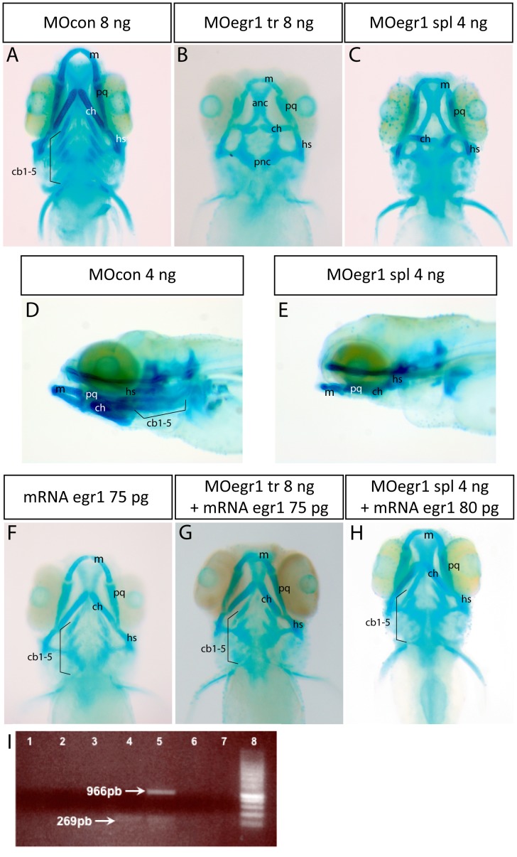
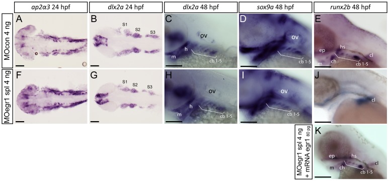
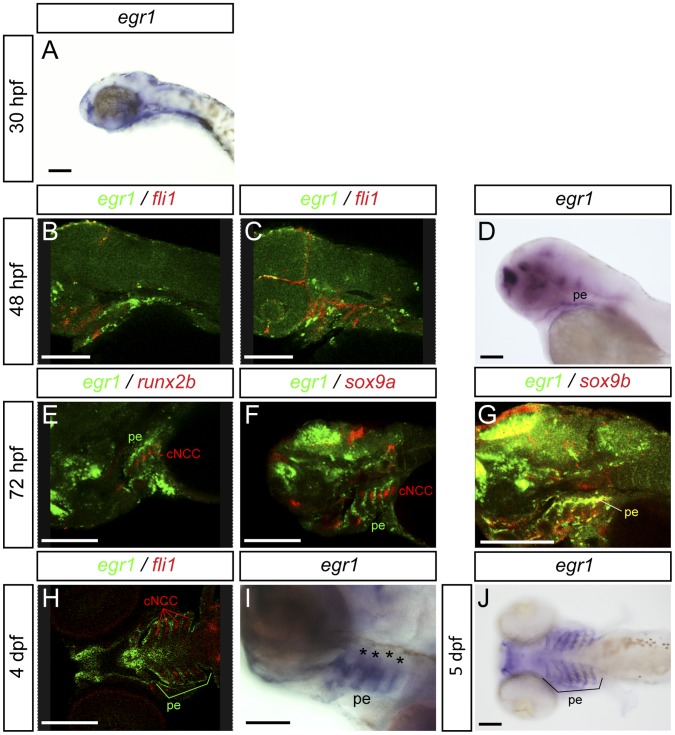
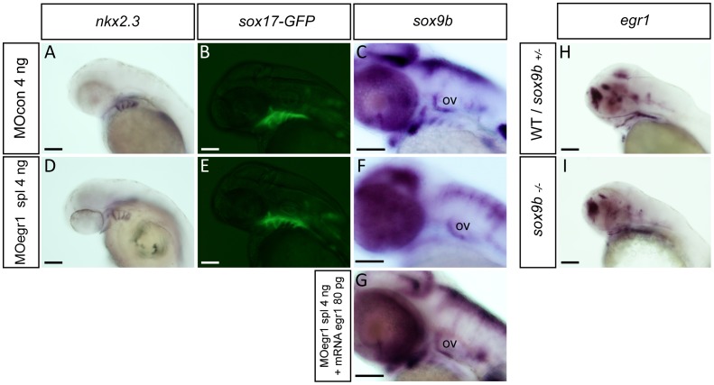
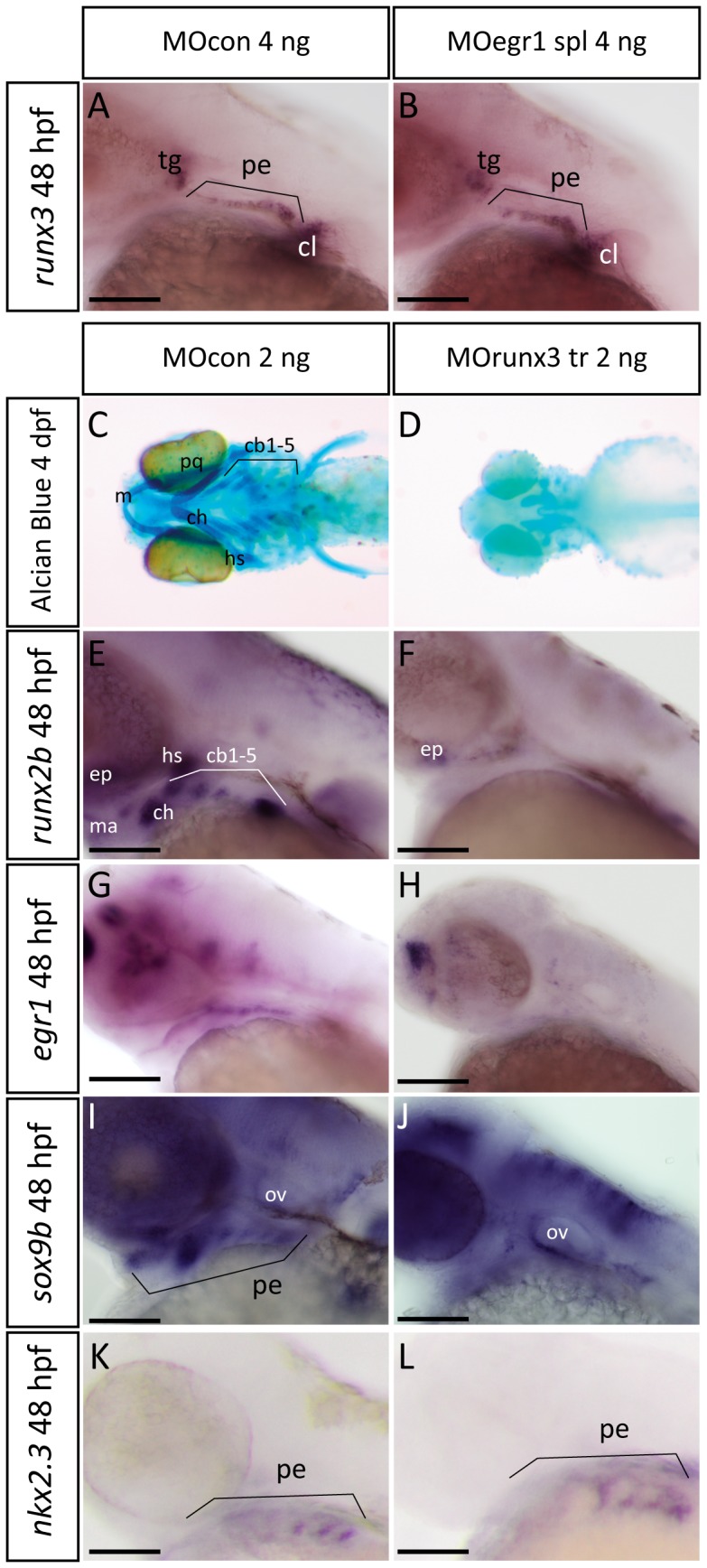
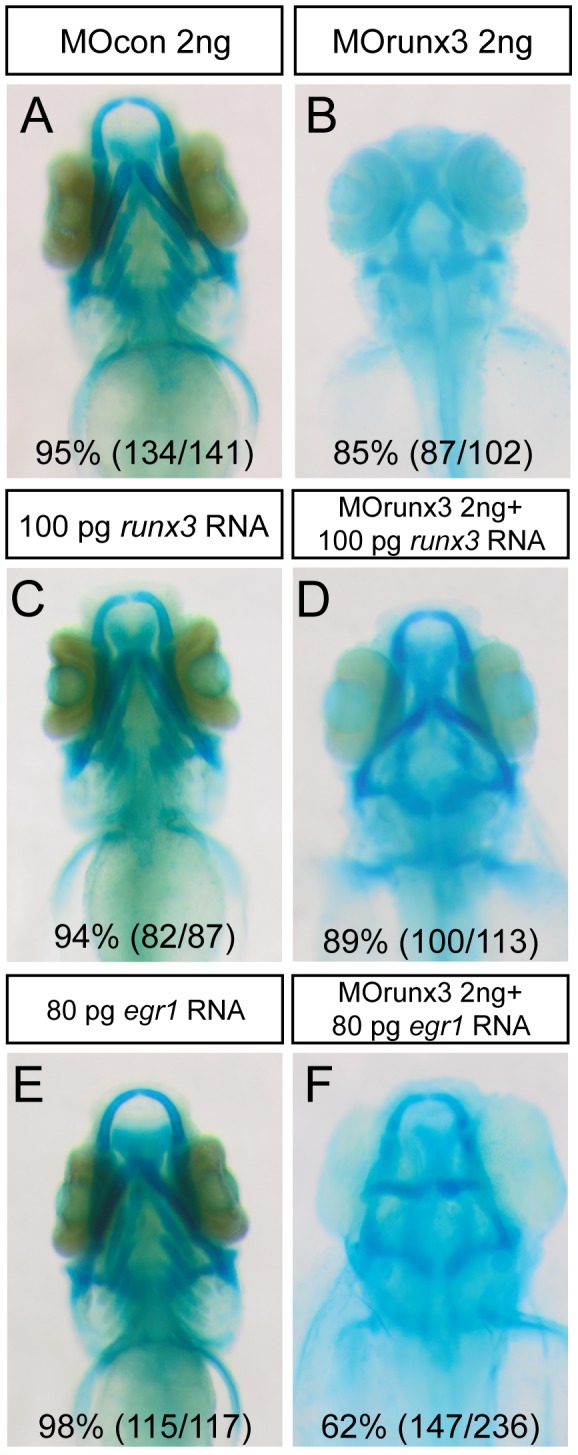
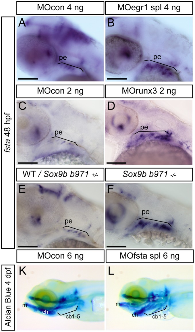
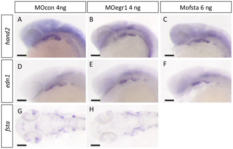
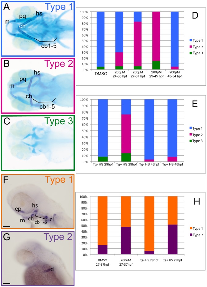
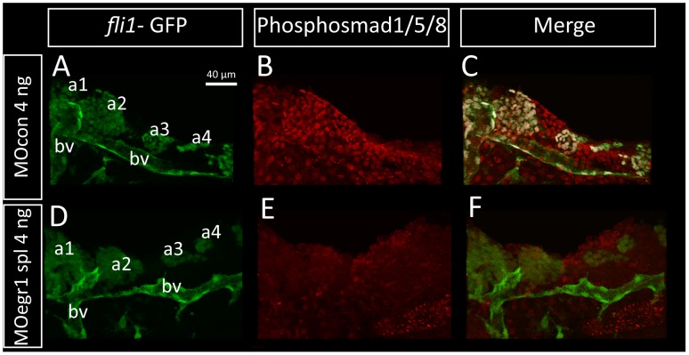
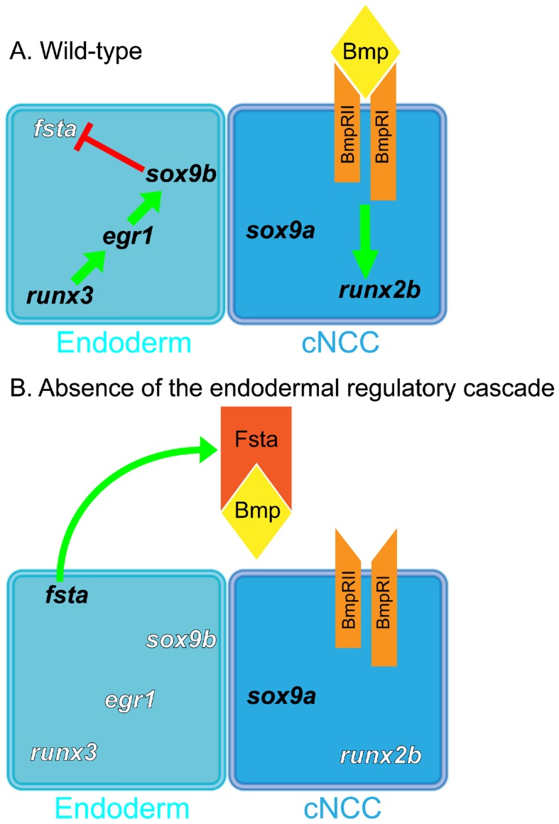
The cartilaginous elements forming the pharyngeal arches of the zebrafish derive from cranial neural crest cells. Their proper differentiation and patterning are regulated by reciprocal interactions between neural crest cells and surrounding endodermal, ectodermal and mesodermal tissues. In this study, we show that the endodermal factors Runx3 and Sox9b form a regulatory cascade with Egr1 resulting in transcriptional repression of the fsta gene, encoding a BMP antagonist, in pharyngeal endoderm. Using a transgenic line expressing a dominant negative BMP receptor or a specific BMP inhibitor (dorsomorphin), we show that BMP signaling is indeed required around 30 hpf in the neural crest cells to allow cell differentiation and proper pharyngeal cartilage formation. Runx3, Egr1, Sox9b and BMP signaling are required for expression of runx2b, one of the key regulator of cranial cartilage maturation and bone formation. Finally, we show that egr1 depletion leads to increased expression of fsta and inhibition of BMP signaling in the pharyngeal region. In conclusion, we show that the successive induction of the transcription factors Runx3, Egr1 and Sox9b constitutes a regulatory cascade that controls expression of Follistatin A in pharyngeal endoderm, the latter modulating BMP signaling in developing cranial cartilage in zebrafish.


Conflict of interest statement

Competing Interests: The authors have declared that no competing interests exist.

Figures

Figure 1
Figure 1. Knock-down of egr1 severely affects head cartilage formation at 4 dpf.
(A–I) Head cartilages were stained with Alcian Blue in morpholino treated larvae at 4 dpf; ventral (A–C) and lateral (D,E) views are shown. (A,D) control 8 ng MOcon treated larvea. (B) 8 ng translation MOegr1 injected larveas display an absence of ceratobranchials and a reduction of size and mis-shaping of pharyngeal cartilage compared to controls (A); (C,E) 4ng splicing MOegr1 injected embryos display similar cartilage defects than 8 ng translation MOegr1 (B). (F) Ectopic expression of Egr1 does not significantly affect cartilage development. (G) Rescue of 8 ng MOegr1 tr treated larvae restores all cartilaginous elements of the viscerocranium. (H) A complete restoration of all cartilage elements is obtained by rescuing 4 ng splicing MOegr1 injected larvae. Meckel’s cartilage (m), palatoquadrate (pq), ceratohyal (ch) and hyosymplectic (hs), ceratobranchials 1 to 5 (cb1-5). (I) Agarose gel electrophoresis analysis of RT-PCR products from mRNA of injected embryos: 1) control mRNA; 2) mRNA of embryos injected with MOcon 4ng and without reverse transcriptase; 3) mRNA of embryos injected with MOegr1 spl 4 ng and without reverse transcriptase; 4) cDNA of embryos injected with MOcon 4 ng. Presence of a band at 269 bp, intron has been spliced properly; 5) cDNA of embryos injected with MOegr1 spl 4 ng. Presence of a band at 966 bp indicating that intron has not been properly spliced. However a residual band at 269 bp reveals that the mRNA has been partially spliced; 6) and 7) cDNA of MOcon 4 ng and MOegr1 spl 4 ng injected embryos that have not undergone the PCR step; 8) molecular weight marker.
Figure 2
Figure 2. Only late chondrogenic and osteogenic marker genes display decreased expression in egr1 morphants between 24 and 48 hpf.
In situ hybridization was performed at the indicated stages for various cartilage markers, lateral views, anterior to the left. Scale bars 100 µm. (A–E) 4 ng MOcon treated control embryos, (F,G,H,I,J) 4ng splicing MOegr1 injected embryos and (K) rescue. (A,F) At 24 hpf, ap2α3 expression in cranial neural crest cells (cNCC) is not altered in morphants. (B,C,G,H) cNCC marker dlx2a is normally expressed in egr1 morphants (G,H) compared to control embryos (B,C) at 24 and 48 hpf. (D,I) Expression of the essential chondrogenic gene sox9a is not changed at 48 hpf by egr1 knock-down. (E,J,K) At 48 hpf, runx2b transcripts are absent in pharyngeal cartilage precursor cells in 4 ng MOegr1 spl embryos. Expression of runx2b is maintained in the cleithrum (cl) and ethmoid plate (ep). (K) Rescue by injection of 80 pg mRNA egr1 restores all runx2b expression domains at 48 hpf. Otic vesicle (ov), mandible (m), ceratohyal (ch), hyosymplectic (hs), ceratobranchial pairs 1 to 5 (cb1-5), cleithrum (cl), ethmoid plate (ep), stream of cNCCs (S1–S3).
Figure 3
Figure 3. Expression of egr1 in the pharyngeal region between 30 hpf to 5 dpf is restricted to endoderm and epithelium.
Lateral (A–G,I) and ventral (H,J) views, anterior to the left. Scale bars 100 µm. Images of double in situ hybridizations were taken by confocal microscopy and pictures of individual Z-sections are shown. (A) egr1 transcripts are observed in the pharyngeal region starting at 30 hpf in endoderm. (B,C) At 48 hpf, double in situ hybridization for egr1 (green) and fli1 (red); egr1 transcripts are localized in pharyngeal endoderm and do not colocalize with fli1 mRNA in pharyngeal cartilage precursor cells. (D) egr1 is expressed in pharyngeal endoderm. (E–G) At 3 dpf, egr1 (green) does not colocalize with runx2b (red) (E) or sox9a (red) (F) in cartilage, while (G) egr1 (green) mRNAs colocalize with those for the pharyngeal endoderm marker sox9b (red). (H) At 4 dpf, egr1 (green) is never expressed in cells in pharyngeal cartilage precursor cells expressing fli1 (red). (I) Expression of egr1 at 4 dpf in pharyngeal endoderm. (J) At 5 dpf, egr1 is still expressed in pharyngeal endoderm (stars) and not in pharyngeal cartilage. Pharyngeal endoderm (pe), cranial neural crest cells (cNCC).
Figure 4
Figure 4. Egr1 is required for expression of sox9b in pharyngeal endoderm.
Endodermal gene expression by in situ hybridization (A,C,D,F,G,H,I) or in living transgenic embryos (B,E) in control embryos (A–C,H), egr1 morphants (D–F), rescued embryos (G) and sox9b mutants (I) at 48 hpf. Lateral views, anterior to the left. Scale bars 100 µm. (A,D) nkx2.3 expression is not altered in 4 ng MOegr1 spl injected embryos. (B,E) In living sox17:GFP transgenic embryos, the transgene is correctly expressed in egr1 morphants. (D,F,G) The endodermal marker sox9b is not expressed in the pharyngeal endoderm in 4 ng MOegr1 spl injected embryos, but its expression is rescued upon co-injection of 80 pg egr1 mRNA and spl 4 ng MOegr1. (H, I) In homozygous sox9b−/− embryos, egr1 transcripts are still observed in the pharyngeal endoderm like in the wild-type or heterozygous sox9b+/− embryos. Pharyngeal endoderm (pe), otic vesicle (ov).
Figure 5
Figure 5. Runx3 is required for pharyngeal egr1 and sox9b expression at 48 hpf.
Lateral views of in situ hybridizations (A,B,E–L) with indicated markers and ventral views of Alcian Blue stained embryos (C,D), anterior to the left. Scale bars 100 µm. (A,B) Endodermal runx3 expression in the pharyngeal region is not altered in 4 ng MOegr1 spl morphants. (C,D) runx3 knock-down using 2 ng MOrunx3 tr leads to total absence of viscerocranium and the anterior neurocranium (D) compared to control (C) embryos. (E,F) runx3 morphants do not express runx2b in pharyngeal cartilage precursor cells. (G,H) runx3 morphants do not express egr1 transcripts in pharyngeal endoderm. (I,J) The endodermal marker sox9b is absent in pharyngeal endoderm when runx3 expression is blocked. (K,L) runx3 knock-down does not affect expression of pharyngeal endodermal marker nkx2.3 at 48 hpf. Trigeminal ganglia (tg), pharyngeal endoderm (pe), cleithrum (cl), Meckel’s cartilage (m), palatoquadrate (pq), hyosymplectic (hs), ceratohyal (hs), ceratobranchials 1 to 5 (cb1-5), ethmoid plate (ep), otic vesivle (ov).
Figure 6
Figure 6. Runx3 depleted embryos can be rescued by runx3 and egr1 mRNA.
(A–F) Head cartilages were stained with Alcian Blue in morpholino treated larvae at 4 dpf; ventral views are shown. (A) MOcon 2 ng injected larvae. (B) MOrunx3 2 ng injected larvae do not develop viscerocranium. (C) 100 pg of runx3 mRNA do not affect 4 dpf old larvae cartilage morphology. (D) Injection of 100 pg runx3 mRNA rescues 89% of MOrunx3 2 ng injected eggs. (E) 80 pg of egr1 mRNA. (F) Injection of 80 pg egr1 mRNA rescues 62% of MOrunx3 2 ng injected eggs.
Figure 7
Figure 7. Expression of fsta is increased in runx3 and egr1 morphants and sox9b mutants.
Lateral views of in situ hybridizations, anterior to the left. Scale bars 100 µm. (A–F) Compared to controls or wild-type embryos, expression of fsta is up-regulated in egr1 morphants (A,B), runx3 morphants (C,D), and homozygous sox9b mutants (E,F) at 48 hpf. pharyngeal endoderm (pe). (G,H) 4 dpf Alcian Blue stained larvae injected with MOcon 6 ng (K) and MOfsta 6ng (L). Knock-down of fsta causes a hyperplasia of the viscerocranium. Meckel’s cartilage (m), ceratohyal (ch), ceratobranchials 1 to 5 (cb1-5).
Figure 8
Figure 8. egr1 and fsta knock-down do not affect ventralisation of cranial neural crest cells.
In situ hybridization was performed at 24 hpf, lateral views, anterior to the left. Scale bars 100 µm. (A,D,G) MOcon 4 ng, (B,E,H) MOegr1 4 ng and (C,F) MOfsta 6 ng. No modification in the expression of markers hand2, edn1 and fsta was observed in MOegr1 4 ng or MOfsta 6 ng injected embryos compared to control.
Figure 9
Figure 9. BMP signaling is required between 27 and 37 hpf for runx2b expression and head cartilage development.
(A–E) Cartilage was stained with Alcian Blue in 4 dpf larvae, ventral views are shown, anterior to the left. (A) Type 1 larvae (blue) display a wild-type morphology, all cartilage elements are present and well shapped. (B) Type 2 larvae (pink) lack ceratobranchials and have mis-shaped Meckel’s cartilage, palatoquadrate, ceratohyal and hyosymplectic. (C) Type 3 larvae (green) display a complete absence of viscerocranium and a reduction of the anterior neurocranium. (D,E) Graphs representing the proportions of the three types of larvae after the indicated treatments. (D) Treatment with the BMP inhibitor dorsomorphin (200 µM) most severely affects head cartilage between 29 and 45 hpf; (DMSO) dimethylsulfoxide. (E) Heat shock treatment of (hsp70l:dnBmpr-GFP)w30 transgenic embryos between 27 hpf and 37 hpf most severely affects pharyngeal cartilage development. (F–H) In situ hybridization for runx2b expression at 48 hpf, lateral views, anterior to the left, scale bars 100 µm. (F) Type 1 embryos (orange) have a normal runx2b expression pattern in all pharyngeal cartilage precursor cells, cleithrum and ethmoid plate. (G) Type 2 embryos (purple), compared to type 1 embryos, only express runx2b in cleithrum and weakly in the ethmoid plate. (H) Graph representing the proportions of the two types of larvae after the indicated treatments. Dorsomorphin treatment of wt embryos and heat shock treatment of (hsp70l:dnBmpr-GFP)w30 between 27 hpf and 37 hpf decreases runx2b expression in pharyngeal cartilage, but not in the cleithrum. (DMSO) dimethylsulfoxide, (Tg+) Transgene expressing embryo, (Tg-) Transgene non-expressing siblings, (HS) heat shock. Meckel’s cartilage (m), palatoquadrate (pq), ceratohyal (ch) and hyosymplectic (hs), ceratobranchials 1 to 5 (cb1-5).
Figure 10
Figure 10. Bmp signaling is down-regulated in egr1 morphants.
Pharyngeal cartilage precursor cells were visualized by immunohistochemistry using anti-GFP antibodies (green) in fli-GFP embryos. Activity of the BMP signaling pathway was assessed using antibodies against phospho-Smad1/5/8 (red) in 32 hpf embryos. Ventral view of pharyngeal arches, scale bar 40 µm. (A–F) Pharyngeal cartilage precursor cells were visualized by immunohistochemistry using anti-GFP antibodies (green) in fli1-GFP embryos. Activity of the BMP signaling pathway was assessed using antibodies against phospho-Smad1/5/8 (red) in 32 hpf embryos. Ventral view of pharyngeal arches, scale bar 40 µm. (A,B,C) 4 ng MOcon injected embryos, (D, E, F) 4 ng MOegr1 spl injected embryos. fli1-GFP embryos express the GFP transgene in cartilage precursors and endothelial cells in control (A) and in egr1 morphants (D). In contrast, phospho-Smad1/5/8 is is clearly down regulated in egr1 morphants (E) compared to control embryos (B). (C,F) Overlay images of the two anti-body signals clearly show that phospho-Smad1/5/8 is present in GFP-epressing cartilage precursor cells in control embryos (C), while no colocalization is observed in egr1 morphants (F). (a1) first arch, (a2) second arch, (a3) third arch, (a4) fourth arch, (bv) blood vessel.
Figure 11
Figure 11. Runx3, Egr1 and Sox9b form a regulatory cascade required to modulate Bmp-signaling during cranial cartilage development in zebrafish.
Signaling model in wild-type embryos (A) and in embryos lacking of endodermal regulatory cascade (B). (A) In wild-type embryos, pharyngeal endoderm expresses a regulatory cascade composed of three transcription factors, Runx3, Egr1 and Sox9b, which down-regulates fsta expression that codes for a Bmp antagonist. This down-regulation of fsta enables Bmp ligands to bind to their heterodimeric receptor (BmpRI and BmpRII) and induce runx2b expression in cranial neural crest cells (cNCC). (B) Embryos lacking of any member of Runx3-Egr1-Sox9b cascade have an over-expression of fsta, which its coding protein is secreted from the endoderm. Antagonist Fsta binds to Bmp ligands and inhibit them to bind to their receptor, having for consequence no Bmp-signaling towards the cNCC and no runx2b expression.

Similar articles

Cited by

References

    1. Schilling TF, Kimmel CB (1994) Segment and cell type lineage restrictions during pharyngeal arch development in the zebrafish embryo. Development 120: 483–494. - PubMed
    1. Knight RD, Javidan Y, Zhang T, Nelson S, Schilling TF (2005) AP2-dependent signals from the ectoderm regulate craniofacial development in the zebrafish embryo. Development 132: 3127–3138. - PubMed
    1. Yanagisawa H, Clouthier DE, Richardson JA, Charite J, Olson EN (2003) Targeted deletion of a branchial arch-specific enhancer reveals a role of dHAND in craniofacial development. Development 130: 1069–1078. - PubMed
    1. Sperber SM, Saxena V, Hatch G, Ekker M (2008) Zebrafish dlx2a contributes to hindbrain neural crest survival, is necessary for differentiation of sensory ganglia and functions with dlx1a in maturation of the arch cartilage elements. Dev Biol 314: 59–70. - PubMed
    1. Yan YL, Willoughby J, Liu D, Crump JG, Wilson C, et al. (2005) A pair of Sox: distinct and overlapping functions of zebrafish sox9 co-orthologs in craniofacial and pectoral fin development. Development 132: 1069–1083. - PubMed

Publication types

MeSH terms