. 2012;7(11):e50180.
doi: 10.1371/journal.pone.0050180. Epub 2012 Nov 27.

A new safety concern for glaucoma treatment demonstrated by mass spectrometry imaging of benzalkonium chloride distribution in the eye, an experimental study in rabbits

Affiliations

A new safety concern for glaucoma treatment demonstrated by mass spectrometry imaging of benzalkonium chloride distribution in the eye, an experimental study in rabbits

Françoise Brignole-Baudouin et al. PLoS One. 2012.

Erratum in

  • PLoS One. 2013;8(1). doi:10.1371/annotation/b97d3c0d-b49e-40e3-a6b4-5155bd9bf3c9

Abstract

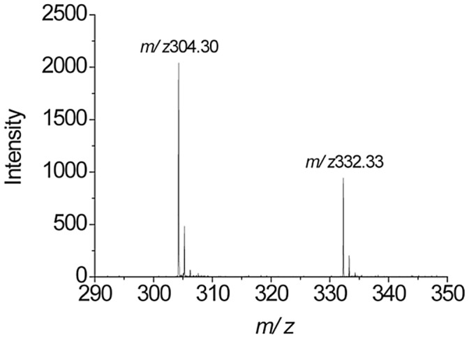
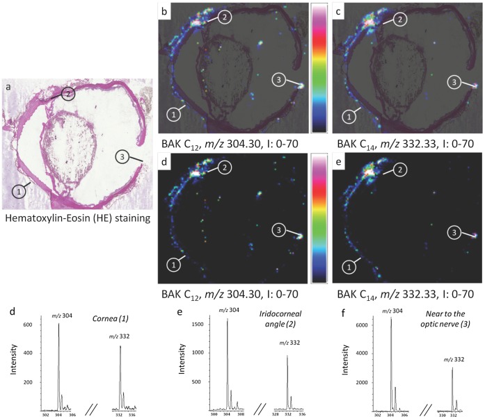
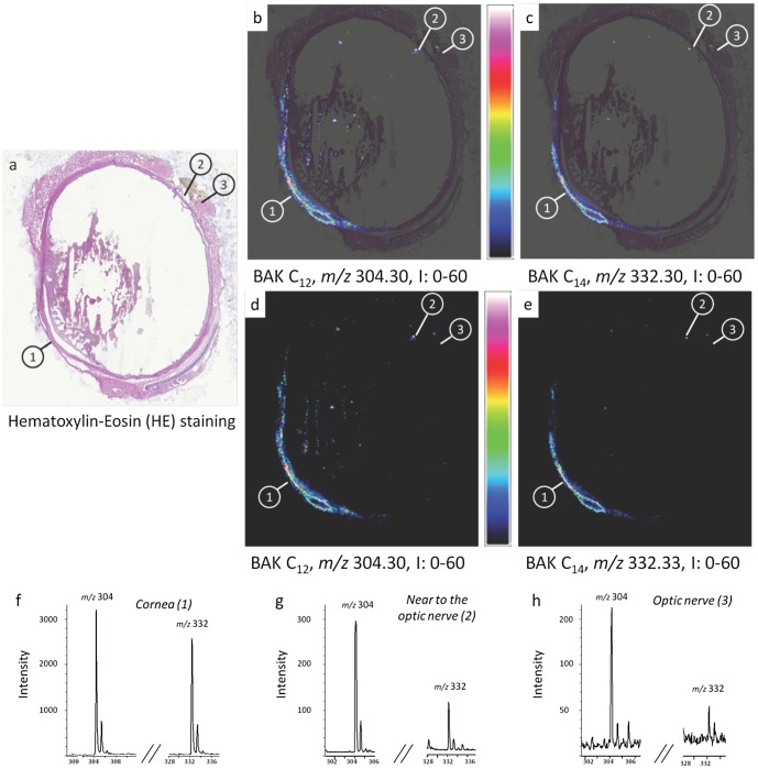
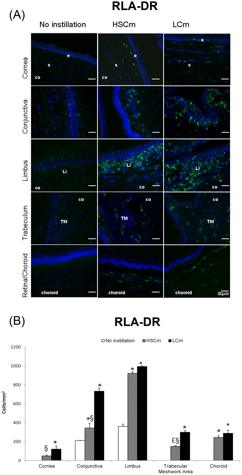
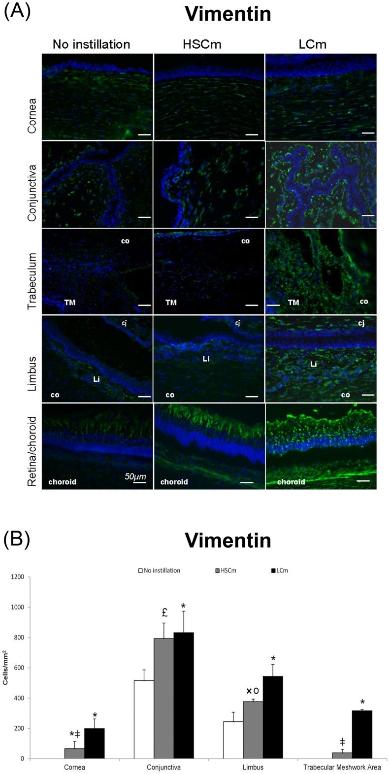
We investigated in a rabbit model, the eye distribution of topically instilled benzalkonium_(BAK) chloride a commonly used preservative in eye drops using mass spectrometry imaging. Three groups of three New Zealand rabbits each were used: a control one without instillation, one receiving 0.01%BAK twice a day for 5 months and one with 0.2%BAK one drop a day for 1 month. After sacrifice, eyes were embedded and frozen in tragacanth gum. Serial cryosections were alternately deposited on glass slides for histological (hematoxylin-eosin staining) and immunohistological controls (CD45, RLA-DR and vimentin for inflammatory cell infiltration as well as vimentin for Müller glial cell activation) and ITO or stainless steel plates for MSI experiments using Matrix-assisted laser desorption ionization time-of-flight. The MSI results were confirmed by a round-robin study on several adjacent sections conducted in two different laboratories using different sample preparation methods, mass spectrometers and data analysis softwares. BAK was shown to penetrate healthy eyes even after a short duration and was not only detected on the ocular surface structures, but also in deeper tissues, especially in sensitive areas involved in glaucoma pathophysiology, such as the trabecular meshwork and the optic nerve areas, as confirmed by images with histological stainings. CD45-, RLA-DR- and vimentin-positive cells increased in treated eyes. Vimentin was found only in the inner layer of retina in normal eyes and increased in all retinal layers in treated eyes, confirming an activation response to a cell stress. This ocular toxicological study confirms the presence of BAK preservative in ocular surface structures as well as in deeper structures involved in glaucoma disease. The inflammatory cell infiltration and Müller glial cell activation confirmed the deleterious effect of BAK. Although these results were obtained in animals, they highlight the importance of the safety-first principle for the treatment of glaucoma patients.


Conflict of interest statement

Competing Interests: The authors acknowledge the fact that three authors are employed by Imabiotech Campus Cité Scientifique, a company specializing in mass spectrometry. These authors made independent analyses that confirmed the results obtained by the other members of the consortium in different laboratories. This does not alter the authors’ adherence to all the PLOS ONE policies on sharing data and materials.

Figures

Figure 1
Figure 1. Chemical structures and MALDI-TOF spectra.
Chemical structure and MALDI-TOF spectrum of benzalkonium homologs used in this study, BAK C12 and BAK C14, are presented with their respective MALDI-TOF spectrum in positive ion mode. The BAK solution instilled in the rabbit eyes contained two thirds of BAK C12 (m/z 304.30) and one third of BAK C14 (m/z 332.33).
Figure 2
Figure 2. MALDI-TOF imaging of whole eye section of a control rabbit.
MALDI-TOF imaging shows the absence of benzalkonium chloride (BAK) in the control eye. (a) Histology image of an adjacent cryosection stained with hematoxylin-eosin (HE) showing three areas of interest: cornea (area 1), nasal iridocorneal angle (area 2) and near to the optic nerve (area 3). (b, c) Overlays between HE and MALDI-TOF images of BAK C12 and C14 eye distributions at m/z 304.32 and 332.36, respectively. Intensities of the ions are represented in colour, based on the intensity scale provided (from black to white). Field of view 18× 6 mm. (d, e, f) MALDI-TOF mass spectra extracted from areas 1, 2 and 3, respectively confirming the absence of BAK C12 and C14.
Figure 3
Figure 3. MALDI-TOF imaging of whole eye section of a rabbit instilled twice a day with one drop of 0.01% benzalkonium chloride (BAK) for 5 months.
MALDI-TOF imaging shows the BAK distribution in a BAK-treated eye. (a) Histology image of an adjacent cryosection stained with hematoxylin-eosin (HE) showing three areas of interest: cornea (area 1), nasal iridocorneal angle (area 2) and optic nerve area (area 3). (b, c) Overlays between HE staining and MALDI-TOF ion images of BAK C12 and C14 distributions in whole eye section at m/z 304.30 and 332.33, respectively. (d, e) MALDI-TOF ion images of BAK C12 and C14 distributions at m/z 304.30 and 332.33, respectively. Intensities of the ions are represented in colour, based on the intensity scale provided (from black to white). Field of view 16×15 mm. (f, g, h) MALDI-TOF mass spectra extracted from areas 1, 2 and 3, respectively, showing BAK C12 and C14 ion peaks.
Figure 4
Figure 4. Round-robin experiment using the AutoFlex speed LRF MALDI-TOF mass spectrometer (ImaBiotech).
MALDI-TOF imaging generated by the AutoFlex speed LRF MALDI-TOF mass spectrometer (ImaBiotech) shows the BAK distribution in whole eye section of a rabbit instilled once a day with one drop of 0.2% benzalkonium chloride (BAK) for 1 month. (a) Histology image of an adjacent cryosection stained with hematoxylin-eosin (HE) showing three areas of interest: cornea (area 1), nasal iridocorneal angle (area 2) and optic nerve area (area 3). (b, c) Overlays between HE staining and MALDI-TOF ion images of BAK C12 and C14 distributions in whole eye section at m/z 304.30 and 332.33, respectively. (d, e) MALDI-TOF ion images of BAK C12 and C14 distributions in whole eye section at m/z 304.30 and m/z 332.33, respectively. Intensities of the ions are represented in colour, based on the intensity scale provided (from black to white). Field of view 8×10 mm. (f, g, h) MALDI-TOF mass spectra extracted from areas 1, 2 and 3, respectively, showing BAK C12 and C14 ion peaks.
Figure 5
Figure 5. Round-robin experiment using the 4800 MALDI-TOF/TOF mass spectrometer (ICSN-CNRS).
MALDI-TOF imaging generated by the 4800 MALDI-TOF/TOF mass spectrometer (ICSN-CNRS) shows the BAK distribution in whole eye section of a rabbit instilled once a day with one drop of 0.2% benzalkonium chloride (BAK) for 1 month. (a) Histology image of an adjacent cryosection stained with hematoxylin-eosin (HE) showing three areas of interest: cornea (area 1), near to optic nerve area (area 2) and optic nerve (area 3). (b, c) Overlays between HE staining and MALDI-TOF ion images of BAK C12 and C14 distributions in whole eye section at m/z 304.32 and m/z 332.36, respectively. (d, e) MALDI-TOF ion images of BAK C12 and C14 distributions in whole eye section at m/z 304.30 and m/z 332.33, respectively, with intensity scale from 0 to 714. Intensities of the ions are represented in color, based on the intensity scale provided (from black to red). Field of view 18×23 mm. (f, g, h) MALDI-TOF mass spectra extracted from areas 1, 2 and 3, respectively, showing BAK C12 and C14 ion peaks.
Figure 6
Figure 6. CD45-positive cell infiltration. (
A) Immunofluorescence staining of leucocytes with CD45 (in green) in rabbit eye cryosections in normal noninstilled rabbit eyes compared with rabbit eye instilled with BAK 0.01% twice a day for 5 months (Low Chronic model, LCm) and 0.2% once a day for 1 month (High Sub-Chronic model, HSCm). Nuclei are stained in blue with DAPI. Scale bar, 50 mm. Cj: conjunctiva; Co: cornea; Li: limbus; s: corneal stroma; e: superficial epithelium; TM: trabecular meshwork. (B) Histogram of CD45 positive cells count (mean cells/mm2±SD) *P<0.001 compared with the normal eye; I P<0.0001 HsCm versus LCm.
Figure 7
Figure 7. RLA-DR-positive cell infiltration.
(A) Immunofluorescence staining of antigen presenting cells expressing RLA-DR (in green) in rabbit eye cryosections in normal non instilled rabbit eyes compared with rabbit eye instilled with BAK 0.01% twice a day for 5 months (Low Chronic model, LCm) and 0.2% once a day for 1 month (High SubChronic model, HSCm). Nuclei in blue are stained with DAPI. Scale bar, 50 mm. Cj: conjunctiva; Co: cornea; Li: limbus; s: corneal stroma; e: superficial epithelium; TM: trabecular meshwork. (B) Histogram of RLA-DR positive cells count (mean cells/mm2±SD) *P<0.001 compared with the normal eye or £ P<0.001 compared with the normal eye; § P<0.01 HsCm versus LCm.
Figure 8
Figure 8. Vimentin-positive cell infiltration.
(A) Immunofluorescence stainings of vimentin (in green) in rabbit eye cryosections in normal non instilled rabbit eyes compared with rabbit eye instilled with BAK 0.01% twice a day for 5 months (Low Chronic model, LCm) and 0.2% once a day for 1 month (High SubChronic model, HSCm). Nuclei in blue are stained with DAPI. Scale bar, 50 mm. Cj: conjunctiva; Co: cornea; Li: limbus; s: corneal stroma; e: superficial epithelium; TM: trabecular meshwork. (B) Histogram of vimentin positive cells count (mean cells/mm2±SD) *P<0.001 compared with the normal eye or £ P<0.001 or × P<0.005 compared with the normal eye; I P<0.0001 or ○ P<0.0001 HsCm versus LCm.

References

    1. Quigley HA (2011) Glaucoma. Lancet 377: 1367–1377. - PubMed
    1. Okabe K, Kimura H, Okabe J, Kato A, Shimizu H, et al. (2005) Effect of benzalkonium chloride on transscleral drug delivery. Invest Ophthalmol Vis Sci 46: 703–8. - PubMed
    1. Chetoni P, Burgalassi S, Monti D, Saettone MF (2003) Ocular toxicity of some corneal penetration enhancers evaluated by electrophysiology measurements on isolated rabbit corneas. Toxicol In Vitro 17: 497–504. - PubMed
    1. The United State Pharmacopoeia 24th rev (2000) The National Formulary 19th ed. Rockville, MD: the United States Pharmacopeial Convention, Inc.
    1. European Pharmacopoeia 7th ed (2010) Council of Europe, Strasbourg.

Publication types

MeSH terms