. 2012;7(11):e50407.
doi: 10.1371/journal.pone.0050407. Epub 2012 Nov 28.

Glutamine treatment attenuates endoplasmic reticulum stress and apoptosis in TNBS-induced colitis

Affiliations

Glutamine treatment attenuates endoplasmic reticulum stress and apoptosis in TNBS-induced colitis

Irene Crespo et al. PLoS One. 2012.

Expression of concern in

Abstract

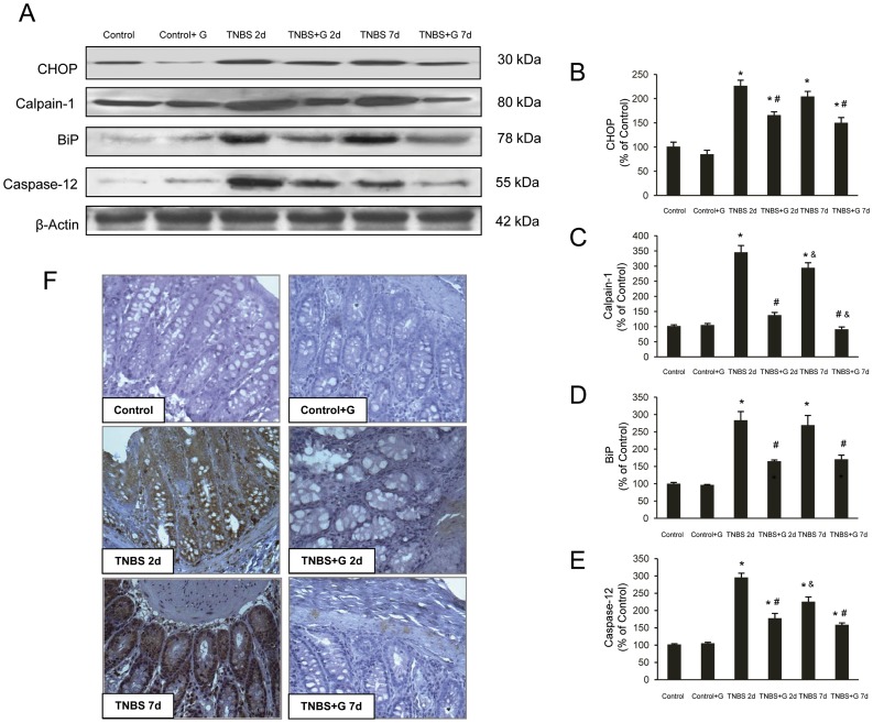
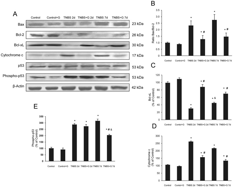
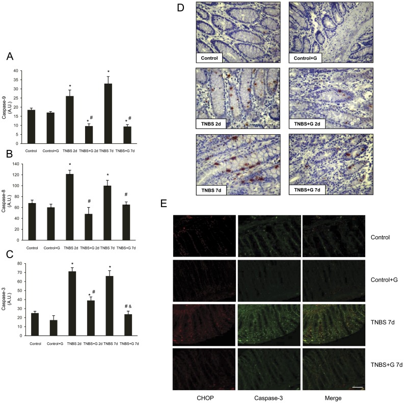
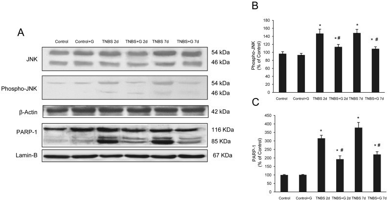
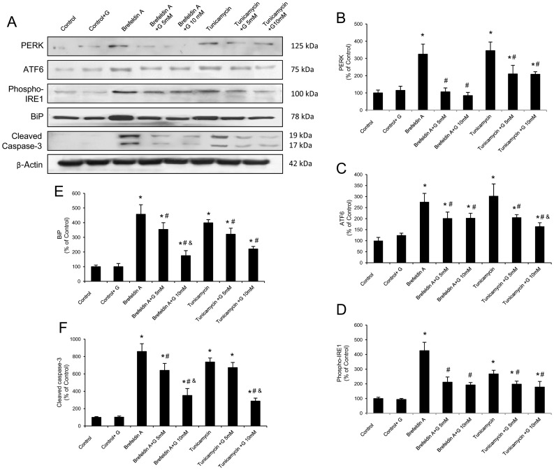
Endoplasmic reticulum (ER) stress and apoptotic cell death play an important role in the pathogenesis and perpetuation of inflammatory bowel disease (IBD). We aimed to explore the potential of glutamine to reduce ER stress and apoptosis in a rat model of experimental IBD. Colitis was induced in male Wistar rats by intracolonic administration of 30 mg of 2,4,6-trinitrobenzene sulfonic acid (TNBS). Glutamine (25 mg/dL) was given by rectal route daily for 2 d or 7 d. Both oxidative stress (TBARS concentration and oxidised/reduced glutathione ratio) and ER stress markers (CHOP, BiP, calpain-1 and caspase-12 expression) increased significantly within 48 h of TNBS instillation, and glutamine attenuated the extent of the changes. Glutamine also inhibited the significant increases of ATF6, ATF4 and spliced XBP-1 mRNA levels induced by TNBS instillation. TNBS-colitis resulted in a significant increase in p53 and cytochrome c expression, and a reduced Bcl-xL expression and Bax/Bcl-2 ratio. These effects were significantly inhibited by glutamine. Treatment with the amino acid also resulted in significant decreases of caspase-9, caspase-8 and caspase-3 activities. Double immunofluorescence staining showed co-localization of CHOP and cleaved caspase-3 in colon sections. Phospho-JNK and PARP-1 expression was also significantly higher in TNBS-treated rats, and treatment with glutamine significantly decreased JNK phosphorylation and PARP-1 proteolysis. To directly address the effect of glutamine on ER stress and apoptosis in epithelial cells, the ER stress inducers brefeldin A and tunicamycin were added to Caco-2 cells that were treated with glutamine (5 mM and 10 mM). The significant enhancement in PERK, ATF6 phosphorylated IRE1, BiP and cleaved caspase-3 expression induced by brefeldin A and tunicamycin was partly prevented by glutamine. Data obtained indicated that modulation of ER stress signalling and anti-apoptotic effects contribute to protection by glutamine against damage in TNBS-induced colitis.


Conflict of interest statement

Competing Interests: The authors have declared that no competing interests exist.

Figures

Figure 1
Figure 1. Glutamine reduces the ER stress induced by TNBS-colitis.
(A–E). Protein from colonic extracts was separated by sodium dodecyl sulfate-polyacrylamide gel electrophoresis, followed by immunoblotting for CHOP, BiP, calpain-1 and caspase-12. CHOP, BiP, calpain-1 and caspase-12 were markedly expressed in rats treated with TNBS alone. However, glutamine administration partially abolished CHOP, BiP, calpain-1 and caspase-12 expression induced by TNBS. Results are representative of four independent experiments. Equal loading of proteins is illustrated by β-actin bands. (A) Representative Western-blot photographs for CHOP, calpain-1, BiP, caspase-12, and β-actin. (B) Densitometric quantification of CHOP. (C) Densitometric quantification of calpain-1. (D) Densitometric quantification of BiP. (E) Densitometric quantification of caspase-12. Data are expressed as mean ± S.E.M. from 8 rats. *P<0.05 compared with control group. #P<0.05 compared with TNBS group. &P<0.05 compared with same group 2 d. (F) Photomicrographs of immunohistochemistry for BiP in sections of colonic samples. Paraffin-embedded sections were immunostained with a BiP antibody. Original magnification: 200X.
Figure 2
Figure 2. Glutamine reduces the apoptotic pathways induced by TNBS-colitis.
(A–E) Protein from colonic extracts was separated by sodium dodecyl sulfate-polyacrylamide gel electrophoresis, followed by immunoblotting for Bax, Bcl-2, Bcl-xL, cytochrome c, p53 and phospho-p53. Bax, cytochrome c and phospho-p53 were markedly expressed in rats treated with TNBS alone. On the contrary, Bcl-2 and Bcl-xL were markedly reduced in rats treated with TNBS alone. However, glutamine administration partially abolished the changes in Bax, Bcl-2, Bcl-xL, cytochrome c and phospho-p53 expression induced by TNBS. Results are representative of four independent experiments. Equal loading of proteins is illustrated by β -actin bands. (A) Representative Western-blot photographs for Bax, Bcl-2, Bcl-xL, cytochrome c, p53, phospho-p53, and β-actin. (B) Densitometric quantification of ratio Bax/Bcl-2. (C) Densitometric quantification of Bcl-xL. (D) Densitometric quantification of cytochrome c. (E) Densitometric quantification of phospho-p53. Data are expressed as mean ± S.E.M. from 8 rats. *P<0.05 compared with control group. #P<0.05 compared with TNBS group. &P<0.05 compared with same group 2 d.
Figure 3
Figure 3. Glutamine reduces the caspase activities induced by TNBS-colitis.
(A–C) Colon activity of caspase-9, caspase-8, and caspase-3 were markedly increased in rats treated with TNBS alone. However, glutamine administration abolished caspase-9, caspase-8, and caspase-3 activities induced by TNBS. (A) Activity of caspase-9. (B) Activity of caspase-8. (C) Activity of caspase-3. Data are expressed as mean ± S.E.M. from 8 rats. *P<0.05 compared with control group. #P<0.05 compared with TNBS group. &P<0.05 compared with same group 2 d. (D) Photomicrographs of immunohistochemistry for cleaved caspase-3 in sections of colonic samples. Paraffin-embedded sections were immunostained with a cleaved caspase-3 antibody. Original magnification: 200X. (E) Photomicrographs of double immunofluorescence for CHOP and cleaved caspase-3 in sections of colonic samples. Paraffin-embedded sections were double staining with a CHOP (red) and cleaved caspase-3 (green) antibodies, and the yellow colour visualized in the merged images represented co-localization of CHOP with cleaved caspase-3. Data shown are representative from four rats. Scale bar 50 µm.
Figure 4
Figure 4. Glutamine reduces the JNK phosphorylation and PARP-1 proteolysis induced by TNBS-colitis.
(A–C) Protein from colonic extracts was separated by sodium dodecyl sulphate-polyacrylamide gel electrophoresis, followed by immunoblotting for JNK, phospho-JNK (cytosolic extracts) and PARP-1 (nuclear extracts). Phospho-JNK and PARP-1 were markedly expressed in rats treated with TNBS alone. However, glutamine administration partially abolished phospho-JNK and PARP-1 expression induced by TNBS. Results are representative of four independent experiments. Equal loading of proteins is illustrated by β-actin (cytosolic extracts) and lamin B (nuclear extracts) bands. (A) Representative Western-blot photographs for JNK, phospho-JNK, β-actin, PARP-1 and lamin-B. (B) Densitometric quantification of phospho-JNK. (C) Densitometric quantification of PARP-1. Data are expressed as mean ± S.E.M. from 8 rats. *P<0.05 compared with control group. #P<0.05 compared with TNBS group.
Figure 5
Figure 5. Glutamine reduces the ER stress and apoptosis induced by brefeldin A and tunicamycin in Caco-2 cells.
(A–F) Protein from Caco-2 cells was separated by sodium dodecyl sulfate-polyacrylamide gel electrophoresis, followed by immunoblotting for PERK, ATF6, phosphorylated IRE1 (phospho-IRE1), BiP and cleaved caspase-3. PERK, ATF6 phospho-IRE1, BiP and cleaved caspase-3 were markedly expressed in cells treated with brefeldin A or tunicamycin alone. However, glutamine administration (5 and 10 mM) partially abolished the changes induced by brefeldin A and tunicamycin. Results are representative of four independent experiments. Equal loading of proteins is illustrated by β-actin bands. (A) Representative Western-blot photographs for PERK, ATF6, phospho-IRE1, BiP, cleaved caspase-3 and β-actin. (B) Densitometric quantification of PERK. (C) Densitometric quantification of ATF6. (D) Densitometric quantification of phospho-IRE1. (E) Densitometric quantification of BiP. (F) Densitometric quantification of cleaved caspase-3. Data are expressed as mean ± S.E.M. *P<0.05 compared with control group. #P<0.05 compared with same stress inducer without glutamine group. &P<0.05 compared with same stress inducer +5 mM glutamine group.

References

    1. Fillmann H, Kretzmann NA, San-Miguel B, Llesuy S, Marroni N, et al. (2007) Glutamine inhibits over-expression of pro-inflammatory genes and down-regulates the nuclear factor kappaB pathway in an experimental model of colitis in the rat. Toxicology 236: 217–226. - PubMed
    1. Kretzmann NA, Fillmann H, Mauriz JL, Marroni CA, Marroni N, et al. (2008) Effects of glutamine on proinflammatory gene expression and activation of nuclear factor kappa B and signal transducers and activators of transcription in TNBS-induced colitis. Inflamm Bowel Dis 14: 1504–1513. - PubMed
    1. Kaser A, Martínez-Naves E, Blumberg RS (2010) Endoplasmic reticulum stress: implications for inflammatory bowel disease pathogenesis. Curr Opin Gastroenterol 26: 318–326. - PMC - PubMed
    1. Kaser A, Blumberg RS (2011) Autophagy, microbial sensing, endoplasmic reticulum stress, and epithelial function in inflammatory bowel disease. Gastroenterology 140: 1738–1747. - PMC - PubMed
    1. Ron D, Walter P (2007) Signal integration in the endoplasmic reticulum unfolded protein response. Nat Rev Mol Cell Biol 8: 519–529. - PubMed

Publication types

MeSH terms