. 2012:2012:638064.
doi: 10.1155/2012/638064. Epub 2012 Nov 13.

Fundus autofluorescence and optical coherence tomography findings in branch retinal vein occlusion

Affiliations

Fundus autofluorescence and optical coherence tomography findings in branch retinal vein occlusion

Tetsuju Sekiryu et al. J Ophthalmol. 2012.

Abstract

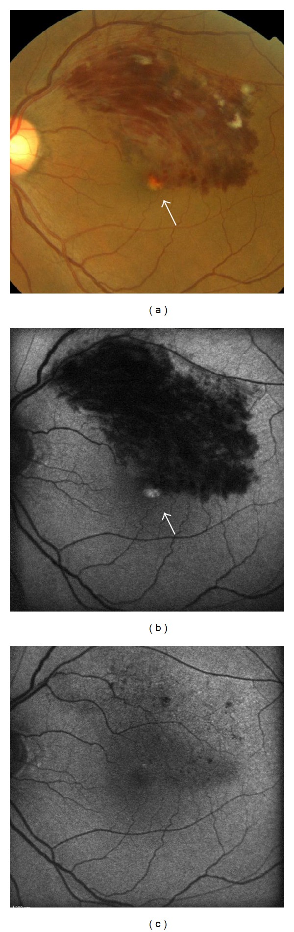
Purpose. To describe the findings of fundus autofluorescence (FAF) and optical coherence tomography (OCT) in patients with branch retinal vein occlusion (BRVO). Methods. In this institutional, retrospective, observational case series, FAF was evaluated in 65 eyes with BRVO in 64 consecutive patients and compared with visual acuity, OCT findings, and other clinical observations. Results. Five types of autofluorescence appeared during the course of BRVO: (1) petaloid-shaped hyperautofluorescence in the area of macular edema and (2) hyperautofluorescence coincident with yellow subretinal deposits. (3) Diffuse hyperautofluorescence appeared within the area of serous retinal detachment (SRD) and OCT showed precipitates on the undersurface of the retina in 5/5 of these eyes (100%). (4) The area of vein occlusion showed diffuse hyperautofluorescence after resolution of the retinal bleeding. (5) Hard exudates exhibited hyper- or hypoautofluorescence. OCT indicated that most of the hard exudates with hyperautofluorescence were located on the retinal pigment epithelium. Conclusions. Hyperautofluorescence associated with subretinal fluid or hard exudate appeared in the subretinal space. This type of hyperautofluorescence may be attributed to blood cell or macrophages. FAF and OCT are noninvasive modalities that provide additional information regarding macular edema due to BRVO.


Figures

Figure 1
Figure 1
Fundus photograph, OCT, fluorescein angiograph, and autofluorescence images for Patient 1 (a 56-year-old man). A color fundus photograph obtained at the first visit showed an upper temporal vein occlusion in the right eye (A). The best-corrected visual acuity in his right eye was 0.3. A “cotton wool” patch was noticed in the affected area. OCT and fluorescein angiography revealed cystoid macular edema (B). Fluorescein angiography showed a petaloid-shaped hyperfluorescence at the fovea (C). Fundus autofluorescence revealed petaloid autofluorescence corresponding to a cystoid space at the fovea (D).
Figure 2
Figure 2
Fundus photograph and fundus autofluorescence images for Patient 5 (a 60-year-old man). Color fundus photograph (a) and fundus autofluorescence (b). Yellowish deposition (white arrow) appeared at the fovea 2 months after the onset of BRVO. Hyperautofluorescence disappeared after resolution of the deposits (c).
Figure 3
Figure 3
Fundus photograph, OCT, and autofluorescence images for Patient 3 (a 56-year-old man). Color fundus photography at the first visit showed a lower temporal vein occlusion in the left eye (A). The dashed line indicates the OCT scanning line. The OCT image (B) showed widespread deposits on the outer surface of the retina (black arrows). An intraretinal deposit was observed in the outer plexiform layer of the retina (white arrow). Fundus autofluorescence (C) at the first visit showed diffuse hyperautofluorescence in the area of serous retinal detachment. The eye was treated with an intravitreal bevacizumab injection. After the serous retinal detachment was resolved, fundus autofluorescence (D) showed hyperautofluorescence in the nasal area of the fovea (arrowhead). The best-corrected visual acuity in the patient's left eye was 0.7 at the final visit.
Figure 4
Figure 4
Fundus autofluorescence and OCT images for Patient 4 (a 67-year-old man). Retinal bleeding in the inferotemporal area was resolved 19 weeks after intravitreal bevacizumab injection. (a1) Fundus autofluorescence showed hyperautofluorescence in the inferotemporal area. (a2) High magnification image of the fovea. Hyperautofluorescence appeared in the corresponding area of cystoid macular edema (white arrowheads). The OCT image (b) showed a defect of the inner/outer segments at the fovea (arrows) and thinning of the retina (black arrowheads) corresponding to the area that showed diffuse hyperautofluorescence after resolution of edema (right). The dashed line indicates the OCT scanning line.
Figure 5
Figure 5
Fundus photograph, fundus autofluorescence, and OCT images for Patient 6 (a 52-year-old woman). The fundus photograph (A) showed a dense retinal hemorrhage and a subretinal hemorrhage. Fundus autofluorescence (B) showed petaloid hyperautofluorescence at the fovea. Extensive exudates were noted 4 months after the first visit (C). Hyperautofluorescence corresponding to the exudates was not observed (D). The color of the exudates in the foveo-papillary area turned yellowish 7 months after the first visit (E). The dashed line indicates the OCT scanning line. Fundus autofluorescence (F) showed hyperautofluorescence in the area corresponding to the yellowish exudates in the foveo-papillary area (white arrows). The exudates in the temporal area (black arrows) showed hypoautofluorescence. OCT (G) at the same visit showed deposits on the retinal pigment epithelium (white arrows) in the foveo-papillary area and intraretinal deposits (white arrows) in the temporal area. The IS/OS disappeared (arrowheads). Fluorescein angiography (H) did not reveal abnormalities in the area corresponding to the area of hyperautofluorescence (white arrowheads).

References

    1. Sperduto RD, Hiller R, Chew E, et al. Risk factors for hemiretinal vein occlusion: comparison with risk factors for central and branch retinal vein occlusion: the eye disease case-control study. Ophthalmology. 1998;105(5):765–771. - PubMed
    1. Risk factors for branch retinal vein occlusion. The Eye Disease Case-control Study Group. American Journal of Ophthalmology. 1993;116(3):286–296. - PubMed
    1. Argon laser photocoagulation for macular edema in branch vein occlusion. The Branch Vein Occlusion Study Group. American Journal of Ophthalmology. 1984;98(3):271–282. - PubMed
    1. Glacet-Bernard A, Coscas G, Chabanel A, Zourdani A, Lelong F, Samama MM. Prognostic factors for retinal vein occlusion: a prospective study of 175 cases. Ophthalmology. 1996;103(4):551–560. - PubMed
    1. Spaide RF, Lee JK, Klancnik JK, Jr., Gross NE. Optical coherence tomography of branch retinal vein occlusion. Retina. 2003;23(3):343–347. - PubMed

LinkOut - more resources