. 2012:2012:139268.
doi: 10.5402/2012/139268. Epub 2012 Nov 14.

General aspects of colorectal cancer

Affiliations

General aspects of colorectal cancer

Josep J Centelles. ISRN Oncol. 2012.

Abstract

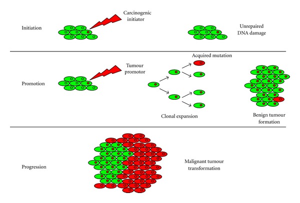
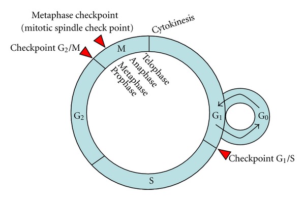
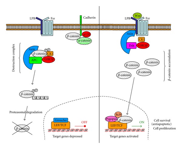
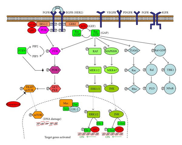
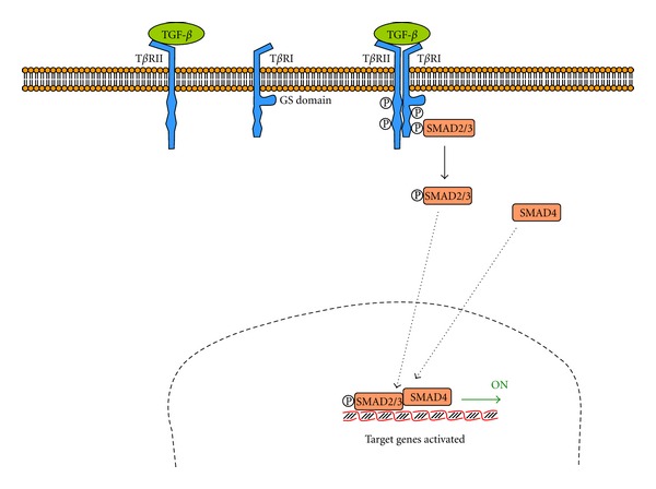
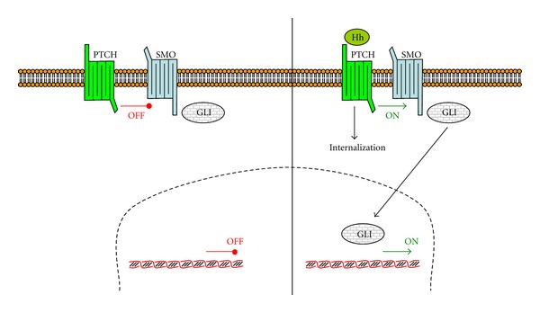
Colorectal cancer (CRC) is one of the main causes of death. Cancer is initiated by several DNA damages, affecting proto-oncogenes, tumour suppressor genes, and DNA repairing genes. The molecular origins of CRC are chromosome instability (CIN), microsatellite instability (MSI), and CpG island methylator phenotype (CIMP). A brief description of types of CRC cancer is presented, including sporadic CRC, hereditary nonpolyposis colorectal cancer (HNPCC) or Lynch syndromes, familiar adenomatous polyposis (FAP), MYH-associated polyposis (MAP), Peutz-Jeghers syndrome (PJS), and juvenile polyposis syndrome (JPS). Some signalling systems for CRC are also described, including Wnt-β-catenin pathway, tyrosine kinase receptors pathway, TGF-β pathway, and Hedgehog pathway. Finally, this paper describes also some CRC treatments.


Figures

Figure 1
Figure 1
Classical theory of carcinogenesis. Cancer is generated by a three-step process: initiation, promotion, and progression. Several mutations are needed to develop a cancer.
Figure 2
Figure 2
Cell cycle and its control. G0 stands for gap 0, G1 for gap 1, S for synthesis, G2 for gap 2, and M for mitosis. Cell cycle includes interface (G1, S, and G2), and M (including prophase, metaphase, anaphase, and telophase). Nondividing cells (G0 or resting phase) are not considered to be in the cell cycle. Cytokinesis follows mitosis. Checkpoint G1/S decides if cells go to rest (G0) or to S. Checkpoint G2/M decides if cells can begin mitosis. Checkpoint M occurs in metaphase of mitosis and it is in charge of biorientation.
Figure 3
Figure 3
Canonical Wnt-β-catenin pathway. Wnt signaling pathway is shown in the “OFF” (left hand side) and “ON” (right hand side) states. In the absence of a Wnt signal, the destruction complex phosphorylates and ubiquinates β-catenin, being therefore destroyed by the proteasome. In the presence of a Wnt signal, as the dishevelled protein (Dsh) recruits the Axin2 and inhibits GSK-3, β-catenin is not phosphorylated and therefore not destroyed. It can translocate to the nucleus and activate transcriptions genes (adapted from [72]).
Figure 4
Figure 4
Tyrosine kinase receptors pathway. Tyrosine kinase receptors include the following dimmers receptors: EGFR, epidermal growth factor receptor (also known as HER2 or ErbB2); HER, human epidermal growth factor receptor; VEGFR, vascular endothelial growth factor receptor; FGFR, fibroblast growth factor receptor; PDGFR, platelet-derived growth factor receptor. IGFR, insulin growth factor receptor, is also a tyrosine kinase receptor. When a growth factor binds to the receptor, a cascade of phosphorylations is initiated, which finishes with activation of transcription genes (adapted from [73]).
Figure 5
Figure 5
Transforming growth factor-β (TGF-β) pathway. TGF-β receptor is a dimer formed by TβRII (with a binding site for TGF-β) and TβRI (with a glycine/serine rich domain). The complete receptor is autophosphorylated and recruits and phosphorylates a gene regulatory protein called SMAD (adapted from [74]).
Figure 6
Figure 6
Hedgehog pathway. In absence of Hedgehog (Hh), PTCH1 binds to SMO, whereas in presence of Hh, the complex Hh-PTCH1 internalizes and releases SMO. Free SMO activates a family of transcription factors, including GLI (adapted from [75]).

References

    1. Howlader N, Noone AM, Krapcho M, et al., editors. SEER Cancer Statistics Review, 1975–2009 (Vintage 2009 Populations) Bethesda, Md, USA: National Cancer Institute; 2012. http://seer.cancer.gov/csr/1975_2009_pops09/
    1. Güller U, Zettl A, Worni M, et al. Molecular investigation of lymph nodes in colon cancer patients using one-step nucleic acid amplification (OSNA). A new road to better staging? Cancer. In press. - PMC - PubMed
    1. Bomme L, Heim S, Bardi G, et al. Allelic imbalance and cytogenetic deletion of 1p in colorectal adenomas: a target region identified between DIS199 and DIS234. Genes Chromosomes Cancer. 1998;21:185–194. - PubMed
    1. Bomme L, Lothe RA, Bardi G, Fenger C, Kronborg O, Heim S. Assessments of clonal composition of colorectal adenomas by fish analysis of chromosomes 1, 7, 13 and 20. International Journal of Cancer. 2001;92(6):816–823. - PubMed
    1. Thorsteinsson M, Kirkeby LT, Hansen R, et al. Gene expression profiles in stages II and III colon cancers: application of a 128-gene signature. International Journal of Colorectal Disease. In press. - PubMed