

The changing phenotype of microglia from homeostasis to disease
- PMID: 23210447
- PMCID: PMC3514090
- DOI: 10.1186/2047-9158-1-9
The changing phenotype of microglia from homeostasis to disease
Abstract
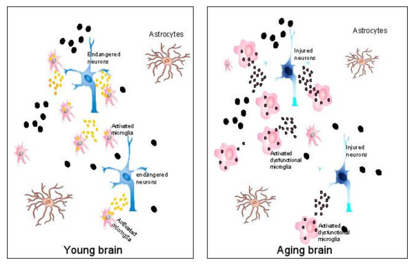
It has been nearly a century since the early description of microglia by Rio-Hortega; since then many more biological and pathological features of microglia have been recognized. Today, microglia are generally considered to be beneficial to homeostasis at the resting state through their abilities to survey the environment and phagocytose debris. However, when activated microglia assume diverse phenotypes ranging from fully inflamed, which involves the release of many pro-inflammatory cytokines, to alternatively activated, releasing anti-inflammatory cytokines or neurotrophins, the consequences to neurons can range from detrimental to supportive. Due to the different experimental sets and conditions, contradictory results have been obtained regarding the controversial question of whether microglia are "good" or "bad." While it is well understood that the dual roles of activated microglia depend on specific situations, the underlying mechanisms have remained largely unclear, and the interpretation of certain findings related to diverse microglial phenotypes continues to be problematic. In this review we discuss the functions of microglia in neuronal survival and neurogenesis, the crosstalk between microglia and surrounding cells, and the potential factors that could influence the eventual manifestation of microglia.
Figures
References
-
- Noda M, Kettenmann H, Wada K. Anti-inflammatory effects of kinins via microglia in the central nervous system. Biol Chem. 2006;387:167–171. - PubMed
LinkOut - more resources
Full Text Sources
Other Literature Sources
Research Materials
