. 2012 Apr 18;1(1):5.
doi: 10.1186/2162-3619-1-5.

Roles of Polo-like kinase 3 in suppressing tumor angiogenesis

Affiliations

Roles of Polo-like kinase 3 in suppressing tumor angiogenesis

Dazhong Xu et al. Exp Hematol Oncol. .

Abstract

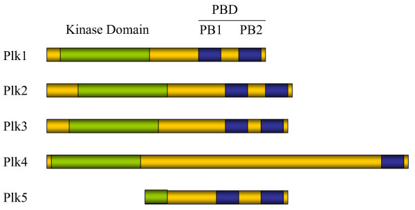
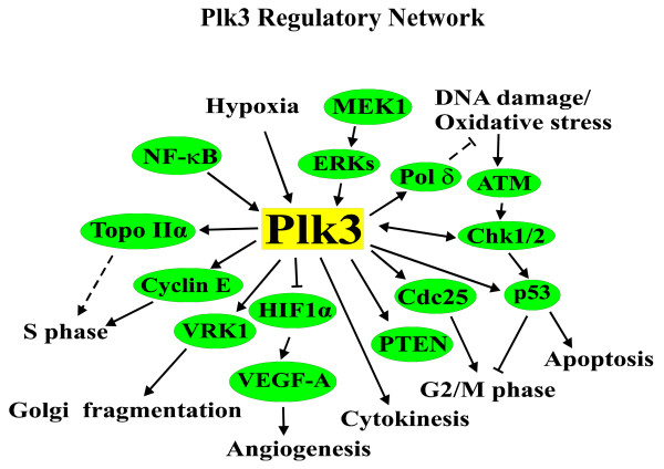
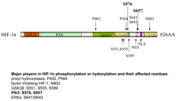
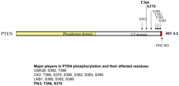
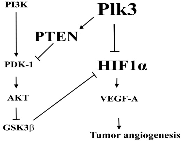
Angiogenesis is essential for promoting growth and metastasis of solid tumors by ensuring blood supply to the tumor mass. Targeting angiogenesis is therefore an attractive approach to therapeutic intervention of cancer. Tumor angiogenesis is a process that is controlled by a complex network of molecular components including sensors, signaling transducers, and effectors, leading to cellular responses under hypoxic conditions. Positioned at the center of this network are the hypoxia-inducible factors (HIFs). HIF-1 is a major transcription factor that consists of two subunits, HIF-1α and HIF-1β. It mediates transcription of a spectrum of gene targets whose products are essential for mounting hypoxic responses. HIF-1α protein level is very low in the normoxic condition but is rapidly elevated under hypoxia. This dramatic change in the cellular HIF-1α level is primarily regulated through the proteosome-mediated degradation process. In the past few years, scientific progress has clearly demonstrated that HIF-1α phosphorylation is mediated by several families of protein kinases including GSK3β and ERKs both of which play crucial roles in the regulation of HIF-1α stability. Recent research progress has identified that Polo-like kinase 3 (Plk3) phosphorylates HIF-1α at two previously unidentified serine residues and that the Plk3-mediated phosphorylation of these residues results in destabilization of HIF-1α. Plk3 has also recently been found to phosphorylate and stabilize PTEN phosphatase, a known regulator of HIF-1α and tumor angiogenesis. Given the success of targeting protein kinases and tumor angiogenesis in anti-cancer therapies, Plk3 could be a potential molecular target for the development of novel and effective therapeutic agents for cancer treatment.


Figures

Figure 1
Figure 1
Domain structures of human Plks. All Plks have a highly conserved kinase domain and a polo box domain (PBD). Plk 1-3 and 5 have two polo boxes (PB) while Plk4 has one discernable PB. Human Plk5 has a truncated kinase domain.
Figure 2
Figure 2
Known functions of Plk3. Plk3 regulates a number of cellular activities by modulating a wide array of cellular components. Arrows indicate activation, T bars indicate inhibition.
Figure 3
Figure 3
Plk3 expression in selected human tumors. Immunohistochemistry was performed on paraffin sections of indicated human tumor and corresponding normal tissue samples using the Plk3 antibody. IgGs were used as control.
Figure 4
Figure 4
Plk3 mRNA and protein expression in selected human melanoma cell lines and tissue samples. A. Real-time PCR was performed on RNA samples from normal melanocytes and selected human melanoma cell lines with DNA primers for Plk3. B. Western blot was performed on normal melanocytes and selected human melanoma cell lines with Plk3 and β-actin antibodies. C. Immunohistochemistry was performed on paraffin sections of human melanoma specimens and corresponding normal skin tissues using the Plk3 antibody.
Figure 5
Figure 5
Domain structure of human HIF-1α, showing Plk3 phosphorylation sites. Human HIF-1α contains a basic helix-loop-helix domain (bHLH), a PAS domain, an oxygen-dependent degradation domain (ODDD), an N-terminal transactivation domain (N-TAD), and a C-terminal transactivation domain (C-TAD). The hydroxylation sites (P402, P564, and N803) are indicated. The Plk3 phosphorylation sites (S576, S657) are bolded. NES: nuclear exporting signal. NLS: nuclear localization signal.
Figure 6
Figure 6
Domain structure of human PTEN, showing Plk3 phosphorylation sites. Human PTEN contains an N-terminal phosphatase domain, a C-terminal C2 domain, and a PDZ binding domain. Major phosphorylation sites at the C-terminal region are indicated. Plk3 phosphorylation sites are bolded.
Figure 7
Figure 7
Proposed model for Plk3 regulation of HIF-1α. Plk3 directly phosphorylates HIF-1α and facilitates its degradation. Plk3 also phosphorylates PTEN and stabilizes it, thereby increasing overall PTEN activity. PTEN inhibits the PI3K pathway and leads to activation of GSK3β, which phosphorylates and destabilize HIF-1α. Inhibition of HIF-1α by Plk3 through direct and indirect mechanisms results in reduced tumor angiogenesis.

Similar articles

Cited by

References

    1. Pugh CW, Ratcliffe PJ. Regulation of angiogenesis by hypoxia: role of the HIF system. Nat Med. 2003;9:677–684. doi: 10.1038/nm0603-677. - DOI - PubMed
    1. Folkman J. Angiogenesis. Annu Rev Med. 2006;57:1–18. doi: 10.1146/annurev.med.57.121304.131306. - DOI - PubMed
    1. Liao D, Johnson RS. Hypoxia: a key regulator of angiogenesis in cancer. Cancer Metastasis Rev. 2007;26:281–290. doi: 10.1007/s10555-007-9066-y. - DOI - PubMed
    1. Folkman J. Tumor angiogenesis: therapeutic implications. N Engl J Med. 1971;285:1182–1186. doi: 10.1056/NEJM197111182852108. - DOI - PubMed
    1. Semenza GL. Targeting HIF-1 for cancer therapy. Nat Rev Cancer. 2003;3:721–732. doi: 10.1038/nrc1187. - DOI - PubMed