. 2013 Feb;344(2):348-59.
doi: 10.1124/jpet.112.197483. Epub 2012 Dec 4.

AT1 receptor antagonism is proangiogenic in the brain: BDNF a novel mediator

Affiliations

AT1 receptor antagonism is proangiogenic in the brain: BDNF a novel mediator

Ahmed Alhusban et al. J Pharmacol Exp Ther. 2013 Feb.

Abstract

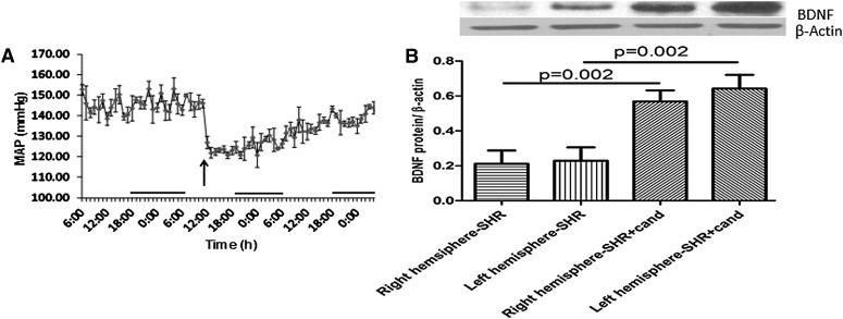
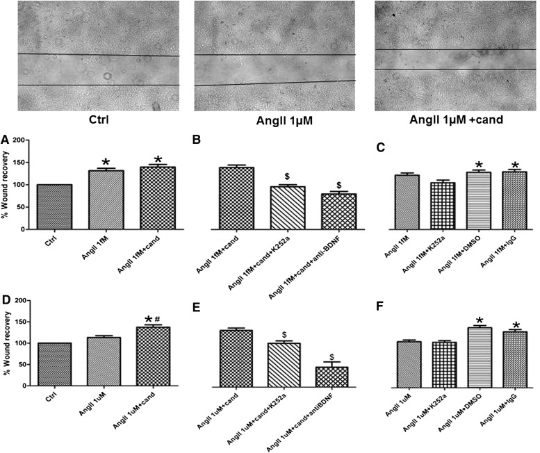
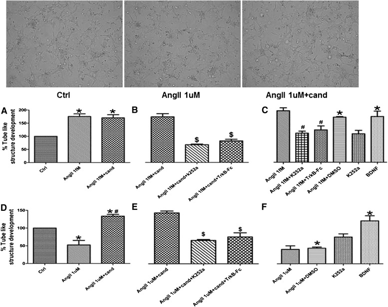
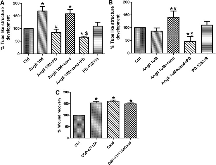
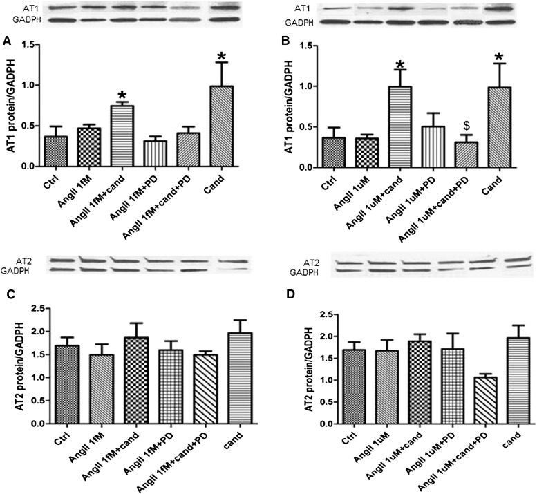
Candesartan is an angiotensin II type 1 receptor blocker (ARB) that has been to shown to limit ischemic stroke and improve stroke outcome. In experimental stroke, candesartan induces a proangiogenic effect that is partly attributable to vascular endothelial growth factor. Brain-derived neurotrophic factor (BDNF) is a member of the neurotrophin family that has been reported to have angiogenic effects and play an important role in recovery after stroke. The purpose of this investigation was to determine the role of BDNF in the proangiogenic effect of candesartan in the brain under hypertensive conditions. Accordingly, spontaneously hypertensive rats were treated with candesartan, and brain tissue samples were collected for quantification of BDNF expression. In addition, human cerebromicrovascular endothelial cells were treated with either low-dose (1 ƒM) or high-dose (1 µM) angiotensin II alone or in combination with candesartan (0.16 µM) to assess the effect of candesartan treatment and BDNF involvement in the behavior of endothelial cells. Candesartan significantly increased the expression of BDNF in the SHR (P < 0.05). In addition, candesartan reversed the antiangiogenic effect of the 1-µM dose of AngII (P = 0.0001). The observed effects of candesartan were ablated by neutralizing the effects of BDNF. Treatment with the AT2 antagonist PD-123319 significantly reduced tube-like formation in endothelial cells. AT2 stimulation induced the BDNF expression and migration (P < 0.05). In conclusion, candesartan exerts a proangiogenic effect on brain microvascular endothelial cells treated with angiotensin II. This response is attributable to increased BDNF expression and is mediated through stimulation of the AT2 receptor.


Figures

Fig. 1.
Fig. 1.
Hypertension and AT1 blockade affects the expression of BDNF. Blood pressure transmitters were implanted intraperitoneally in SHRs. Animals had sham surgery and received a single dose of candesartan (0.3 mg/kg), and the mean arterial blood pressure was monitored (A). Arrow indicates time of candesartan administration; n = 6. SHRs underwent sham surgery and were randomized to receive either candesartan (0.3 mg/kg) or saline intravenously (6 per group); 24 hours later, the animals were sacrificed and the brains were extracted. Right and left hemispheres were separated and processed for immunoblotting (B).
Fig. 2.
Fig. 2.
Angiotensin II and AT1 blockade affects the expression of BDNF and the angiogenic potential in hCMECs. hCMECs were cultured to confluence, followed by serum starvation for 16 hours. Cells were treated with a concentration range of AngII for different periods. The expression of BDNF was assessed using immunoblotting (A) n = 3–5. hCMECs were treated with AngII (1ƒM or 1µM) for 6 hours, followed by treatment with candesartan (0.16 µM) for 10 hours, and the expression of BDNF was assessed (B) n = 3. The ability of candesartan to modulate the angiogenic response of hCMECs was evaluated using in vitro matrigel tube formation assay (C) and wound recovery assay (D) in response to treatment with a concentration range of candesartan (0–1.6 µM) n = 3. (A and B) Data presented as mean ± S.E.M., * P < 0.05. (C and D) *Significantly different from control; $Significantly different from candesartan (0.16 µM). Overall, P = 0.0014, F=8.19 (C) and P < 0.0001, F=22.44 (D).
Fig. 3.
Fig. 3.
AngII modulates the proliferation of hCMECs. Proliferative response of hCMECs was evaluated using BrdU incorporation assay. hCMECs were treated with AngII (1ƒM or 1µM) for 6 hours, followed by candesartan (0.16 µM) alone or in combination with other treatments. The plates were then processed according to the manufacturer recommendations. Data are presented as mean ± S.E.M. of three different experiments each in triplicate. *Significantly different from control, #significantly different from AngII in the same group, $significantly different from AngII+cand in the same group; overall, P = 0.005, F=8.85 for AngII 10−9 and P < 0.0001, F=34.97 for AngII 1µM
Fig. 4.
Fig. 4.
AngII modulates the migration of hCMECs. Insert; representative image of control, AngII 1 µM and AngII+candesartan showing candesartan induced migration of hCMECs. hCMECs were cultured to confluence, followed by 10 hours serum starvation. Cells were treated with either AngII 1ƒM or 1µM for 6 hours, and then a scratch was introduced in the monolayer. Cells were then incubated with AngII 1ƒM or 1µM alone or with candesartan (A and D). The involvement of BDNF was assessed using a number of inhibitors for BDNF functions (B and E), which were added to the media 30 minutes before AngII treatment. Data are presented as mean ± S.E.M. of three different experiments each in triplicate. *Significantly different from control, #significantly different from AngII in the same group, $significantly different from AngII+cand in the same group; overall, P < 0.0001, F=16.08 for AngII 1 ƒM and P < 0.0001, F=22.08 for AngII 1 µM.
Fig. 5.
Fig. 5.
AngII modulates the angiogenic potential of hCMECs. Insert: representative images of in vitro tube formation (arrows) showing reduced tube formation rate in AngII 1 µM treated hCMECs and the reversal of AngII 1 µM antiangiogenic effect by candesartan. hCMECS (2 × 10⁴ cells/well)were suspended in a 30:60 solution of matrigel and serum-free media. Angiogenic response of hCMECs to AngII 1 ƒM (A) and 1 µM (D) in the presence and absence of candesartan was evaluated 24 hours after treatment. The involvement of BDNF was assessed through using K252a (Trk inhbitor) or TrkB-Fc (soluble chimeric receptor) (B and E). Data are presented as mean ± S.E.M. of three different experiments each in triplicate. *Significantly different from control, #significantly different from AngII in the same group, $significantly different from AngII+cand in the same group; overall, P < 0.0001, F=12.74 for AngII 1ƒM and P < 0.0001, F=9.54 for AngII 1µM.
Fig. 6.
Fig. 6.
AT2 receptor mediates the angiogenic response in hCMECs. The involvement of AT2 receptor in the angiogenic response to AngII 1 ƒM alone or in combination with candesartan (A) and to the combination of AngII 1 µM and candesartan (B) was assessed using the AT2 antagonist PD-123319 (0.1 µM). The angiogenic response was evaluated using matrigel tube formation assay as described in the methods section. Data are presented as mean ± S.E.M. of three different experiments each in triplicate. *Significantly different from control, #significantly different from AngII in the same group, $significantly different from AngII+cand in the same group; overall, P < 0.0001, F=8.779 for both AngII 1ƒM and AngII 1µM.
Fig. 7.
Fig. 7.
Candesartan induced BDNF expression is mediated through AT2 receptor. To evaluate the involvement of AT2 receptor in candesartan-induced BDNF expression, hCMECs were pretreated with PD-123319 or vehicle, followed by AngII (1 ƒM or 1 µM) in the presence or absence of candesartan (0.16 µM) (A). To further confirm the role of AT2, hCMECs were treated with CGP-42112A (0.1 µM) or vehicle for 16 hours (B). *Significantly different from control, #significantly different from AngII in the same group, $significantly different from AngII+cand in the same group; overall, P = 0.02, F=3.086, n = 4–6.
Fig. 8.
Fig. 8.
AT1 antagonism affects the expression of AT1 receptor in an AT2 receptor–mediated manner. To assess the expression of AT1 (A and B) and AT2 (C and D) receptors in response to the different treatments used. cells were incubated with PD-123319 (0.1 µM) or vehicle for 30 minutes, followed by 6 hours of AngII 1 ƒM (A and C) or 1 µM (B and D). After 6 hours of AngII treatment, cells were coincubated with candesartan or vehicle for 10 hours. Receptor expression was assessed using immunoblotting. Data are presented as mean ± S.E.M.; n = 3–5. *Significantly different from control, #significantly different from AngII in the same group, $significantly different from AngII+cand in the same group; overall, P < 0.0042, F=8.742 for AngII 1 µM.
Fig. 9.
Fig. 9.
AT1 antagonism modulates the phosphorylation of GSK-3β in an AT2 receptor–mediated manner. To assess the phosphorylation of GSK-3β at the inhibitory serine 9 residue, hCMECs were treated as described for AngII receptors expression evaluation. (A) Response to AngII 1 ƒM. (B) AngII 1 µM response. Data are presented as mean ± S.E.M.; n = 3–5. *Significantly different from control, #significantly different from AngII in the same group, $significantly different from AngII+cand in the same group; overall, P = 0.0038, F=6.94 for AngII 1 µM.
Fig. 10.
Fig. 10.
A schematic representation of the results. By blocking AT1 receptors ARBs induce an unopposed stimulation of AT2 receptors. AT2 stimulation induces the expression of BDNF, which will bind to its TrkB receptor to promote a proangiogenic state in hCMECs.

Similar articles

Cited by

References

    1. Dahlöf B, Devereux RB, Kjeldsen SE, Julius S, Beevers G, de, Faire U, Fyhrquist F, Ibsen H, Kristiansson K, Lederballe-Pedersen O, Lindholm LH, Nieminen MS, Omvik P, Oparil S, Wedel H; LIFE Study Group. (2002) Cardiovascular morbidity and mortality in the Losartan Intervention For Endpoint reduction in hypertension study (LIFE): a randomised trial against atenolol. Lancet 359:995–1002. - PubMed
    1. Buharalioglu CK, Song CY, Yaghini FA, Ghafoor HU, Motiwala M, Adris T, Estes AM, Malik KU. (2011) Angiotensin II-induced process of angiogenesis is mediated by spleen tyrosine kinase via VEGF receptor-1 phosphorylation. Am J Physiol Heart Circ Physiol 301:H1043–H1055 - PMC - PubMed
    1. Caporali A, Emanueli C. (2009) Cardiovascular actions of neurotrophins. Physiol Rev 89:279–308 - PMC - PubMed
    1. Chan SH, Wu CW, Chang AY, Hsu KS, Chan JY. (2010) Transcriptional upregulation of brain-derived neurotrophic factor in rostral ventrolateral medulla by angiotensin II: significance in superoxide homeostasis and neural regulation of arterial pressure. Circ Res 107:1127–1139 - PubMed
    1. De Paolis P, Porcellini A, Gigante B, Giliberti R, Lombardi A, Savoia C, Rubattu S, Volpe M. (1999) Modulation of the AT2 subtype receptor gene activation and expression by the AT1 receptor in endothelial cells. J Hypertens 17:1873–1877 - PubMed

Publication types

MeSH terms